. 2011 Feb;178(2):803-16.
doi: 10.1016/j.ajpath.2010.10.034.

Accelerated human mutant tau aggregation by knocking out murine tau in a transgenic mouse model

Affiliations

Accelerated human mutant tau aggregation by knocking out murine tau in a transgenic mouse model

Kunie Ando et al. Am J Pathol. 2011 Feb.

Abstract

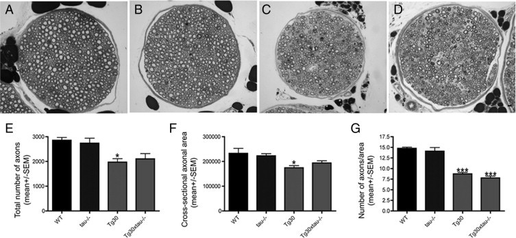
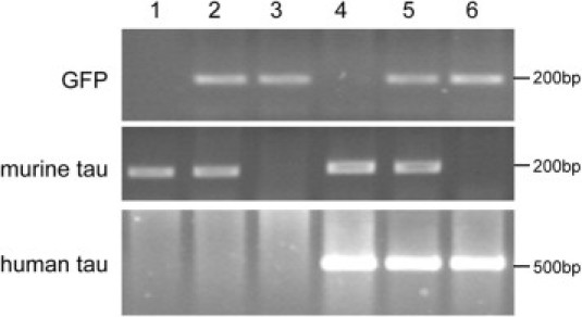
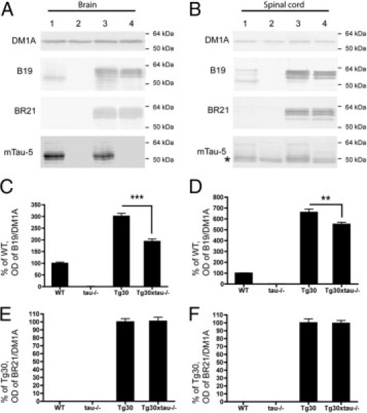
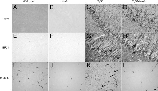
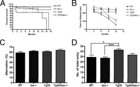
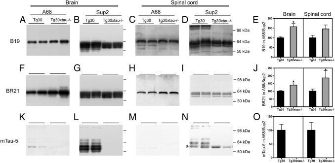
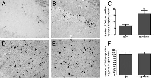
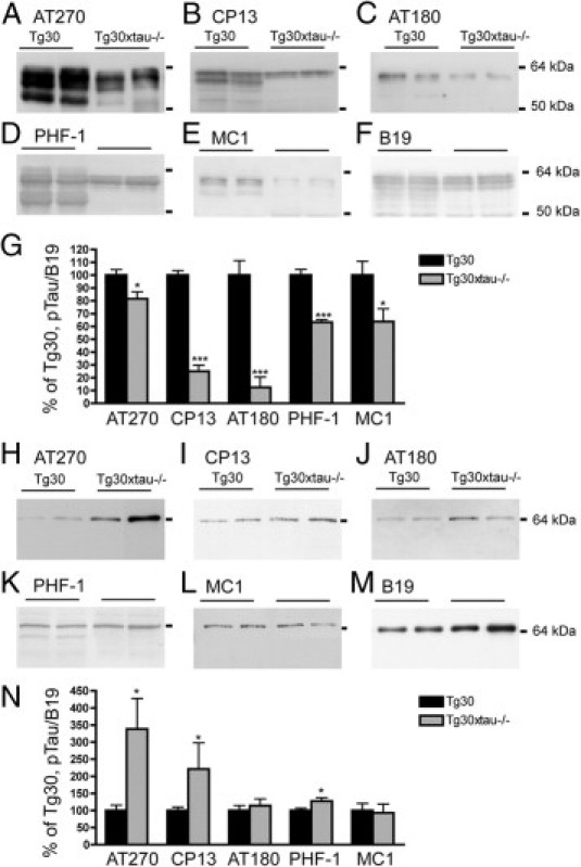
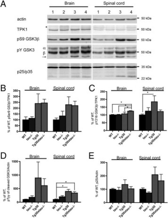
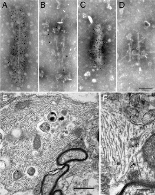
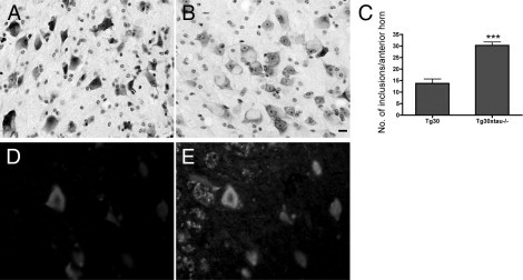
Many models of human tauopathies have been generated in mice by expression of a human mutant tau with maintained expression of mouse endogenous tau. Because murine tau might interfere with the toxic effects of human mutant tau, we generated a model in which a pathogenic human tau protein is expressed in the absence of wild-type tau protein, with the aim of facilitating the study of the pathogenic role of the mutant tau and to reproduce more faithfully a human tauopathy. The Tg30 line is a tau transgenic mouse model overexpressing human 1N4R double-mutant tau (P301S and G272V) that develops Alzheimer's disease-like neurofibrillary tangles in an age-dependent manner. By crossing Tg30 mice with mice invalidated for their endogenous tau gene, we obtained Tg30xtau(-/-) mice that express only exogenous human double-mutant 1N4R tau. Although Tg30xtau(-/-) mice express less tau protein compared with Tg30, they exhibit signs of decreased survival, increased proportion of sarkosyl-insoluble tau in the brain and in the spinal cord, increased number of Gallyas-positive neurofibrillary tangles in the hippocampus, increased number of inclusions in the spinal cord, and a more severe motor phenotype. Deletion of murine tau accelerated tau aggregation during aging of this mutant tau transgenic model, suggesting that murine tau could interfere with the development of tau pathology in transgenic models of human tauopathies.


Figures

Supplemental Figure s1
Semi-thin cross-section of the sciatic nerve in wild-type (A), tau−/− (B), Tg30 (C), and Tg30xtau−/− mice (D). The total number of axons (E), mean cross-sectional area (F), and density of axons (G) were similar in wild-type and tau−/− mice, but reduced in Tg30 and Tg30xtau−/− mice. *P <0.05 and *** P <0.001 by one-way ANOVA with Bonferroni post hoc tests (n = 3 for wild-type; n = 4 for tau−/−; n = 6 for Tg30 and Tg30xtau−/−). Scale bar, 50 μm (A–D).
Figure 1
Figure 1
PCR genotyping of mice resulting from the crossing of Tg30xtau+/− mice with tau+/− mice. PCR amplification was performed with primers for GFP, murine tau, and human tau. Each lane represents a different genotype: lane 1, wild-type; lane 2, tau+/−; lane 3, tau−/−, lane 4, Tg30; lane 5, Tg30xtau+/−; and lane 6, g30Tg30xtau−/−. Only Tg30xtau−/− mice show amplification products for the human tau and GFP transgenes without amplification products of the murine tau gene. Molecular weight markers are indicated on the right.
Figure 2
Figure 2
A, B: Levels of expression of tau proteins in wild-type mice (lane 1), in tau−/− mice (lane 2), in Tg30 mice (lane 3), and in Tg30xtau−/− mice (lane 4). Expression of tau was investigated in brain (A) and in spinal cord (B) with the B19 tau antibody (recognizing both human and mouse tau), the BR21 tau antibody (recognizing only human tau), and the mTau-5 antibody (recognizing only mouse tau). The asterisk in panel B indicates a nonspecific band of mouse immunoglobulin heavy chain detected by the secondary antibody in the soluble fraction of the spinal cord. Human tau was expressed only in Tg30 and Tg30xtau−/− mice and murine tau only in wild-type and Tg30 mice. C, D: The total level of tau (normalized to the level of α-tubulin detected with the DM1A antibody) was significantly decreased in the brain and in the spinal cord of Tg30xtau−/− mice, compared with Tg30 mice. E, F: The level of human tau (normalized to the level of a-tubulin) was not significantly different between Tg30 and Tg30xtau−/− mice. **P < 0.01 and ***P < 0.001 by one-way analysis of variance with Bonferroni post hoc tests [wild-type (WT), n = 4; tau−/−, n = 4; Tg30, n = 6; Tg30xtau−/−, n = 8].
Figure 3
Figure 3
Tau immunohistochemical labeling of tissue sections of the hippocampus of 9-month-old wild-type, tau−/−, Tg30, and Tg30xtau−/− mice. A–D: The B19 tau antibody (reacting with mouse and human tau) shows the somatodendritic accumulation of tau in the CA1 sector of Tg30 and Tg30xtau−/− mouse hippocampus. E–H: The BR21 antibody specific for human tau also shows the somatodendritic accumulation of tau in Tg30 and in Tg30xtau−/− mice and does not show any immunoreactivity in wild-type mice. Some neurons containing neurofibrillary tangles show a stronger labeling in the CA1 sector of Tg30 and Tg30xtau−/− mouse hippocampus. I–L: The mTau-5 antibody specific for murine tau shows an abnormal somatodendritic accumulation of murine tau (arrows) in the subiculum of Tg30 mice, but not in the other genotypes. Blood vessels show a nonspecific labeling due to the use of an anti-mouse secondary antibody. Scale bar = 20 μm (A–L).
Figure 4
Figure 4
Survival and behavioural analysis of wild-type, tau−/−, Tg30 and Tg30xtau−/− mice. A: Kaplan-Meir survival curves. Tg30xtau−/− mice have a significantly reduced survival by comparison with other genotypes and an accelerated mortality during the first 3 months (P < 0.05, by log-rank test, using pairwise multiple comparison methods) (Bonferroni-corrected threshold method) (n = 178 for four genotypes). B: Rotarod testing. Tg30xtau−/− mice developed a motor deficit starting at 3 months and exhibited a more severe motor deficit than other genotypes (n > 11 for each age, for each genotype). C: Y maze test for alternations at 3 to 6 months. The percentage of alternations was not significantly different among all genotypes (wild-type: n = 11; tau−/−: n = 12; Tg30: n = 15; Tg30xtau−/−: n = 16). D: Number of entries in arms during Y-maze test at 3 to 6 months. The total number of entries was similar among genotypes, except that Tg30 mice had a higher number of entries than wild-type and tau−/− mice. *P < 0.05; ***P < 0.001 by one-way analysis of variance with Bonferonni post hoc tests.
Figure 5
Figure 5
The proportion of sarkosyl-insoluble tau was increased in Tg30xtau−/− mice, compared with Tg30 mice. The levels of tau in the sarkosyl-insoluble (A68) and the soluble (Sup2) fractions were analyzed by immunoblotting in the brain and the spinal cord of Tg30 and Tg30xtau−/− mice. The panels show two representative blots for each condition. A–E: Panspecific B19 tau antibody. F–J: Human-specific tau antibody BR21. K–O: Mouse-specific tau antibody mTau5. The B19 and the BR21 antibodies recognize a major 64-kDa insoluble tau species in the brain (A and F) and the spinal cord (C and H) of Tg30 and Tg30xtau−/− mice. Additional minor bands in the sarkosyl-insoluble fraction were detected by the B19 antibody but not by the human-specific BR21 tau antibody. These minor bands are detected by the murine-specific tau antibody (K and M). The proportion of insoluble tau (ratio of insoluble tau to soluble tau) was increased in brain and spinal cord of Tg30xtau−/− mice, as estimated with the B19 antibody (E) and the human-specific BR21 antibody (J). The 110-kDa murine tau isoform (big tau) was detected only in Tg30 mice (D, M, and N). The asterisk in panel N indicates a nonspecific band of mouse immunoglobulin heavy chain detected by the secondary antibody in the soluble fraction of spinal cord (Tg30, n = 5; Tg30xtau−/−, n = 7). *P < 0.05 by unpaired Student's t-test.
Figure 6
Figure 6
The load of neurofibrillary tangles estimated with Gallyas staining was increased in Tg30xtau−/− mice, compared with Tg30 mice (12-month-old mice). A–C: Hippocampus (CA1 sector). The density of NFTs was significantly higher in Tg30xtau−/− than in Tg30 mice (C). *P <0.05 by unpaired t-test (P = 0.032) (Tg30, n = 5; Tg30xtau−/−, n = 6). D–F: Lumbar spinal cord, anterior horn. The density of NFTs was not significantly different between Tg30xtau−/− andTg30 mice (F) (P = 0.8670). The density of NFTs is expressed as the number of NFTs per slice counted in the hippocampus and subiculum in three adjacent sagittal sections (Tg30, n = 6; Tg30xtau−/−, n = 8).
Figure 7
Figure 7
Highly phosphorylated soluble tau was rapidly aggregated into sarkosyl-insoluble fraction in Tg30xtau−/− mouse brain. The phosphorylation of tau in the sarkosyl-soluble fraction S2 (A–F) and sarkosyl-insoluble A68 fraction (H–M) was analyzed by immunoblotting with phosphotau antibodies AT270 (A and H), CP13 (B and I), AT180 (C and J), PHF-1 (D and K), and the conformational antibodies MC1 (E and L) and the polyclonal tau antibody B19 (F and M). The mean relative levels of each phosphotau species estimated by densitometry analysis (normalized to total tau estimated with the B19 antibody) are shown in panels G and N. The majority of pathological phosphotau species were reduced in sarkosyl-soluble fraction, whereas the levels of certain phosphotau species were increased in sarkosyl-insoluble fraction of Tg30xtau−/− mice. *P < 0.05 and ***P < 0.001 by Student's t-test (Tg30, n = 5; Tg30xtau−/−, n = 7).
Figure 8
Figure 8
Kinase activation was analyzed by Western blotting for GSK-3 and p35. A: Immunoblotting for actin, TPK1, pSer9 GSK-3β, pTyr GSK-3, and p25/p35 in brain and spinal cord of wild-type mice (lane 1), tau−/− mice (lane 2), Tg30 mice (lane 3), and Tg30xtau−/− mice (lane 4). The asterisk at TPK1 indicates nonspecific bands of mouse IgG heavy chain. The arrow points to the calpain-cleaved GSK-3. B: The level of pSer9 GSK-3β normalized to TPK1 was generally (but not significantly) increased in the presence of tau pathology. C: The level of pTyr 216 GSK-3β normalized to TPK1 was significantly increased in the brain of Tg30xtau−/− mice and in the spinal cord of Tg30. D: pTyr GSK-3-positive calpain-cleaved GSK-3 was significantly increased in spinal cord of Tg30 and Tg30xtau−/−. E: The level of p25 normalized to actin was generally (but not significantly) increased in Tg30 mice (wild-type, n = 4; tau−/−, n = 4; Tg30, n = 4; Tg30xtau−/−, n = 6).
Figure 9
Figure 9
Electron microscopy of PHF-tau in Tg30 and Tg30xtau−/− mice. A–D: Immunolabeling in electron microscopy of abnormal filaments present in the sarkosyl-insoluble fraction of Tg30 (A and B) and Tg30xtau−/− (C and D) mice. A, C: Anti-human tau antibody (BR21). A strong labeling was observed both in Tg30 and in Tg30xtau−/− mice. B, D: Anti-mouse tau antibody (mTau5). A weak labeling was observed in filaments from Tg30 mice but was absent in Tg30xtau−/− mice. Scale bar = 50 nm (A–D). E, F: Ultrastructural aspects of abnormal filaments in Tg30xtau−/− mice (subiculum). E: A dilated neurite contains bundles of 19-nm straight filaments, admixed with degraded organelles and lysosomal vacuoles. Scale bar = 0.5 μm. F: Under higher magnification, straight filaments and a PHF-like filament (arrow) can be identified. Scale bar = 0.25 μm.
Figure 10
Figure 10
Neurofilament-positive neuronal inclusions in spinal cord were more frequent in Tg30xtau−/− mice than in Tg30 mice. A, B: Nissl staining for hyaline inclusions in the anterior horn of the lumbar spinal cord in Tg30 (A) and Tg30xtau−/− (B) mice. C: The density of inclusions in the anterior horn was significantly increased in Tg30xtau−/− mice. ***P < 0.001 by Student's t-test (n = 5 mice for each genotype). D–E: Double immunolabeling of the anterior horn of Tg30xtau−/− mouse with the PHF-1 tau antibody (D) and a neurofilament antibody (E). Most of the neuronal inclusions were positive for both phosphotau and neurofilament. Scale bar = 20 μm.

References

    1. Selkoe D.J. The origins of Alzheimer disease—A is for amyloid. JAMA. 2000;283:1615–1617. - PubMed
    1. Ballatore C., Lee V.M.Y., Trojanowski J.Q. Tau-mediated neurodegeneration in Alzheimer's disease and related disorders. Nat Rev Neurosci. 2007;8:663–672. - PubMed
    1. van Swieten J., Spillantini M.G. Hereditary frontotemporal dementia caused by Tau gene mutations. Brain Pathol. 2007;17:63–73. - PMC - PubMed
    1. Denk F., Wade-Martins R. Knock-out and transgenic mouse models of tauopathies. Neurobiol Aging. 2009;30:1–13. - PMC - PubMed
    1. Lewis J., Dickson D.W., Lin W.L., Chisholm L., Corral A., Jones G., Yen S.H., Sahara N., Skipper L., Yager D., Eckman C., Hardy J., Hutton M., McGowan E. Enhanced neurofibrillary degeneration in transgenic mice expressing mutant tau and APP. Science. 2001;293:1487–1491. - PubMed

Publication types

MeSH terms