. 2011 Feb;178(2):817-27.
doi: 10.1016/j.ajpath.2010.10.037.

Ophthalmic pterygium: a stem cell disorder with premalignant features

Affiliations

Ophthalmic pterygium: a stem cell disorder with premalignant features

Jeanie Chui et al. Am J Pathol. 2011 Feb.

Abstract

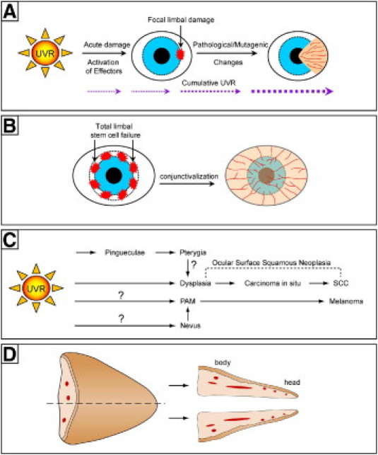
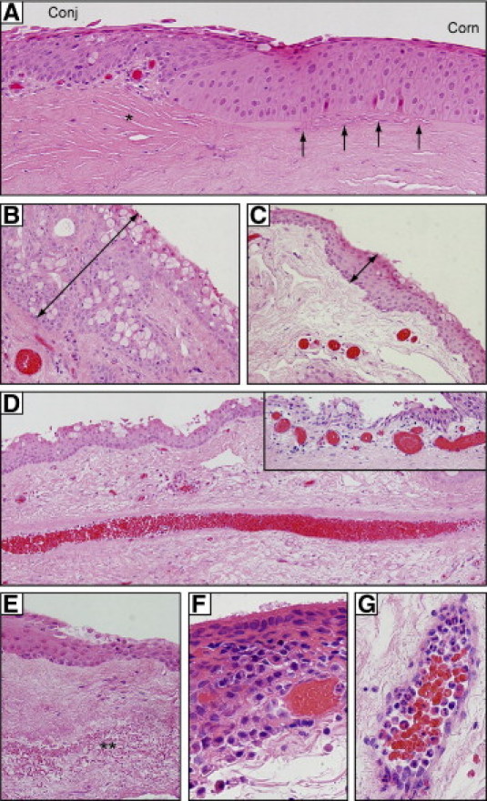
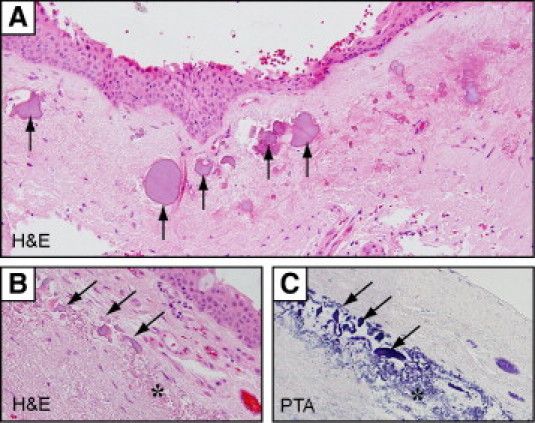
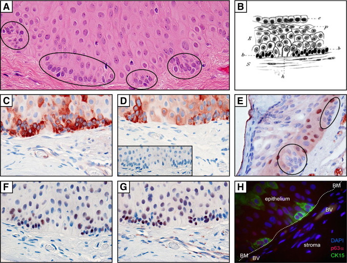
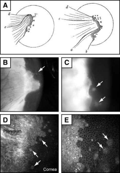
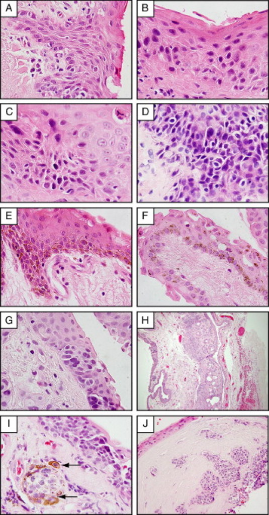
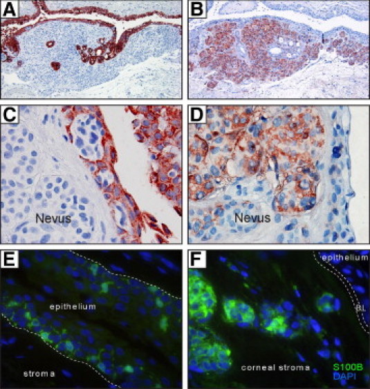
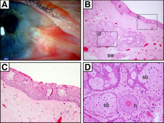
Pterygia are common ocular surface lesions thought to originate from limbal stem cells altered by chronic UV exposure. Traditionally regarded as a degenerative condition, pterygia also display tumor-like features, such as a propensity to invade normal tissue and high recurrence rates following resection, and may coexist with secondary premalignant lesions. This study was initiated to determine the rate of concurrent ocular surface diseases in patients with pterygia recruited from the practice of a single surgeon operating in a Sydney metropolitan hospital. One hundred pterygium specimens were histopathologically reviewed and selected cases were immunohistochemically assessed to confirm diagnosis. Along with previously documented typical features including epithelial proliferation, goblet cell hyperplasia, angiogenesis, inflammation, elastosis, stromal plaques, and Bowman's membrane dissolution, we identified five cases of ocular surface squamous neoplasia, six cases of primary acquired melanosis, two compound nevi (one suspect invasive melanoma), and one dermoid-like lesion. In 18 specimens, clusters of basal epithelial cells that coexpressed cytokeratin-15/-19 and p63-α were identified at the head of the pterygium, coinciding with clinical observation of Fuchs' flecks. Our data show that significant preneoplastic lesions may be associated with pterygium and that all excised pterygia should undergo histological examination. The presence of p63-α-positive epithelial cell clusters supports the hypothesis that pterygia develop from limbal epithelial progenitors.


Figures

Figure 1
Figure 1
The role of cumulative UV radiation exposure in pterygium development. A: Model for the pathogenesis of pterygium: focal limbal damage from UV radiation triggers migration of altered LSCs toward the central cornea. B: In total LSC deficiency, damage to the limbal niche or depletion of stem cell reserves results in conjunctivalization of the cornea from all directions. C: Model of how ocular surface squamous neoplasia and melanoma might arise from pterygia. Question marks with pathways indicate absence of direct supporting clinical or experimental evidence. D: Bisection and orientation of pterygium specimens as assessed in the current study.
Figure 2
Figure 2
Typical histological features of pterygia. A: In the advancing head of a pterygium, conjunctival-like epithelium (Conj) merges abruptly into corneal epithelium (Corn). The underlying Bowman's layer (arrows) is fragmented and precedes a fibrovascular stroma (asterisk). B, C: Goblet cell hyperplasia is apparent in pterygium (B), compared with donor-matched conjunctiva (C). Note the thickness of the epithelial layer (double-headed arrows in B and C). D: Prominent central feeder vessel; inset shows dilated subepithelial vessels. E: Elastotic changes (double asterisk) in pterygium stroma. F: Inflammatory infiltrates in the epithelium. G: Stromal vessels loaded with polymorphonuclear leukocytes. All sections were stained with H&E. Original magnification: ×200 (A and D); ×400 (B, C, D inset, and E); ×1000 oil emersion (F and G).
Figure 3
Figure 3
Stromal plaques in pterygia stained with H&E (A and B) or phosphotungstic acid (PTA) (C). Irregular-shaped stromal plaques (arrows) with an amorphous appearance were frequently associated with elastotic changes (asterisk). Plaques appear lilac in H&E-stained sections and deep blue in PTA-stained sections. Elastin fibers also stained blue with PTA. Original magnification: ×200 (A); ×600 (B and C).
Figure 4
Figure 4
Stem cell microclusters in pterygium tissue. A: H&E-stained section of a pterygium illustrating multiple mini-aggregates of basal epithelial cells (ovals). B: Pterygium epithelium as illustrated by Ernst Fuchs in 1892. C–G: Pterygium immunolabeled with CK-15, CK-19, Ki-67, p63 (pan), and p63α (C–G, respectively), using an indirect immunoperoxidase method with 3-amino-9-ethylcarbazole chromogen; red denotes positive labeling. Staining is absent in sections incubated with control IgG (D, inset). H: Indirect immunofluorescent double-labeling of pterygium epithelial cell clusters with CK-15 (green), p63α (red), and counterstained with DAPI (blue); note basement membrane (BM, indicated by dotted line) and blood vessels (BV). Original magnification, ×1000 oil emersion (all photomicrographs). Image B is reproduced with permission from Springer (original publication: Fuchs E. Ueber der Pterygium. Graefes Archiv Ophthalmol 1892, 38:1–89).
Figure 5
Figure 5
Clinical appearance of Fuchs' flecks in pterygia. A: Original illustrations by Ernst Fuchs show small spots or fleckchen in the cap region at the head of pterygia. B–E: Pterygia with Fuchs' flecks (arrows) under slit lamp (B and C) and in vivo confocal microscopy (D and E). Slit lamp photographs were taken with an iPIX camera (Designs For Vision, Ronkonkoma, NY) and confocal micrographs with an HRT 3 Rostock cornea module (Heidelberg Engineering, Heidelberg, Germany). Confocal images were taken with a 63× objective. The field of view is 400µm × 400µm for (D) and 300µm × 300µm for (E). Image A is reproduced with permission from Springer (original publication: Fuchs E. Ueber der Pterygium. Graefes Archiv Ophthalmol 1892, 38:1–89).
Figure 6
Figure 6
Atypical histological features in pterygia included pterygium epithelium demonstrating papillary folding (A) and dysplastic changes ranging from mild (B) to moderate (C) to severe (D). Melanocytic lesions included racial pigmentation (note layers of pigmented basal melanocytes) (E), PAM without atypia (F), PAM with atypia (G), subconjunctival nonpigmented compound nevus (H), and a nonpigmented nevus (I) from a patient with epidermolysis bullosa that invaded the corneal stroma (J). Arrows in I point to melanophages that have acquied pigmented granules and surround a melanocytic nest. Sections were assessed after H&E staining. Original magnification: ×1000 oil emersion (A–G and I); ×100 (H); ×400 (J).
Figure 7
Figure 7
Cytokeratin and S100B staining of nevi in pterygia. Indirect immunoperoxidase (A–D) and immunofluorescence (E and F) techniques demonstrate CK-19 immunoreactivity in pterygium epithelium (A and C) or S100B immunoreactivity in nevus melanocytes (B, D, and F) and in dendritiform cells scattered within the epithelium (E). In panels A–D, positive labeling is denoted by red color (from 3-amino-9-ethylcarbazole chromogen) and nuclei counterstaining in blue (hematoxylin). In panels E and F, S100B expression is denoted by green immunofluorescence and DAPI counterstaining in blue. In E, the hatched line indicates the pterygium basement membrane; in F, it indicates Bowman's layer (BL). Original magnification: ×100 (A and B); ×1000 oil emersion (C–F).
Figure 8
Figure 8
Cutaneous elements in a recurrent pterygium. Clinical image of a pterygium with hairs growing from the body (A) and its corresponding H&E-stained paraffin sections (B–D). The regions encompassed by the rectangles (c and d) in panel B are magnified in panels C and D, respectively. Goblet cell clusters within the conjunctival portion of the pterygium (C) overlie cutaneous elements (D) such as sebaceous glands (sb), hair follicles (h), and sweat glands (sw). Original magnification: ×100 (B); ×400 (C and D).

References

    1. Duke-Elder S., editor. Diseases of the Outer Eye Part 1: System of Ophthalmology 8. Kimpton; London: 1965. pp. 569–585.
    1. Di Girolamo N., Chui J., Coroneo M.T., Wakefield D. Pathogenesis of pterygia: role of cytokines, growth factors, and matrix metalloproteinases. Prog Retin Eye Res. 2004;23:195–228. - PubMed
    1. Di Girolamo N., McCluskey P., Lloyd A., Coroneo M.T., Wakefield D. Expression of MMPs and TIMPs in human pterygia and cultured pterygium epithelial cells. Invest Ophthalmol Vis Sci. 2000;41:671–679. - PubMed
    1. Di Girolamo N., Coroneo M.T., Wakefield D. Active matrilysin (MMP-7) in human pterygia: potential role in angiogenesis. Invest Ophthalmol Vis Sci. 2001;42:1963–1968. - PubMed
    1. Di Girolamo N., Kumar R.K., Coroneo M.T., Wakefield D. UVB-mediated induction of interleukin-6 and -8 in pterygia and cultured human pterygium epithelial cells. Invest Ophthalmol Vis Sci. 2002;43:3430–3437. - PubMed

Publication types

MeSH terms