. 2011 Feb;178(2):828-37.
doi: 10.1016/j.ajpath.2010.10.026.

Isolation and characterization of progenitor-like cells from human renal proximal tubules

Affiliations

Isolation and characterization of progenitor-like cells from human renal proximal tubules

David Lindgren et al. Am J Pathol. 2011 Feb.

Abstract

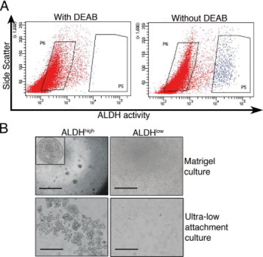
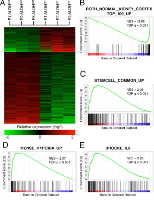
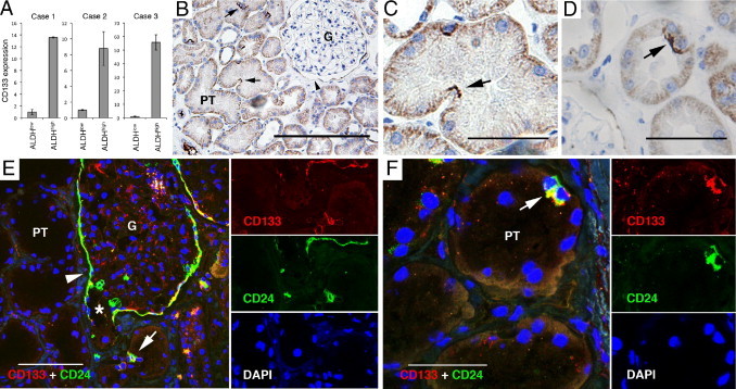
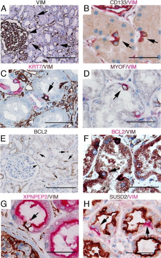
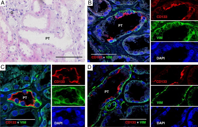
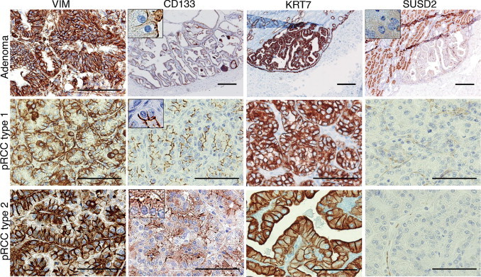
The tubules of the kidney display a remarkable capacity for self-renewal on damage. Whether this regeneration is mediated by dedifferentiating surviving cells or, as recently suggested, by stem cells has not been unequivocally settled. Herein, we demonstrate that aldehyde dehydrogenase (ALDH) activity may be used for isolation of cells with progenitor characteristics from adult human renal cortical tissue. Gene expression profiling of the isolated ALDH(high) and ALDH(low) cell fractions followed by immunohistochemical interrogation of renal tissues enabled us to delineate a tentative progenitor cell population scattered through the proximal tubules (PTs). These cells expressed CD24 and CD133, previously described markers for renal progenitors of Bowman's capsule. Furthermore, we show that the PT cells, and the glomerular progenitors, are positive for KRT7, KRT19, BCL2, and vimentin. In addition, tubular epithelium regenerating on acute tubular necrosis displayed long stretches of CD133(+)/VIM(+) cells, further substantiating that these cells may represent a progenitor cell population. Furthermore, a potential association of these progenitor cells with papillary renal cell carcinoma was discovered. Taken together, our data demonstrate the presence of a previously unappreciated subset of the PT cells that may be endowed with a more robust phenotype, allowing increased resistance to acute renal injury, enabling rapid repopulation of the tubules.


Figures

Figure 1
Figure 1
Isolation of primary renal cortex cells based on ALDH activity. A: Representative FACS analysis of a single-cell suspension of normal renal cortical tissue incubated with fluorescent ALDH substrate. The left panel shows the cellular fluorescence in the presence of the ALDH inhibitor diethylaminobenzaldehyde (DEAB), and the right panel shows the uninhibited reaction with the gate defining the ALDHhigh cells. The FACS gatings used are marked by boxes. In all the experiments, cells were first viability sorted by incubation in 7-amino-actinomycin D. B: The ALDHhigh population was enriched for cells with colony-forming capacity as assessed by plating in Matrigel culture (top row; scale bar = 1000 μm). Inset: A cellular sphere formed in the ALDHhigh culture at higher magnification (×40). The ALDHhigh population was also enriched for cells with the capacity to grow under ultra-low attachment conditions (bottom row; scale bar = 200 μm). The ALDHlow cells displayed none of these capacities.
Figure 2
Figure 2
Gene expression profiling of three pairs of ALDHhigh and ALDHlow cell fractions. A: Gene expression heatmap illustrating the 1445 probes that showed a more than twofold difference between the ALDHhigh and its corresponding ALDHlow populations in all three replicate pairs. B–E: A GSEA of a gene list in which genes were ranked according to their relative expression difference in ALDHhigh compared with ALDHlow cells. Graphs display enrichment score (y axis) versus gene rank (x axis); genes with high relative expression in the ALDHhigh population are given a low rank order value (leftmost tail), and genes with high relative expression in ALDHlow cells are given a high rank order value (rightmost tail) of the gene rank representation. The rank of each gene in the respective gene set is indicated as a horizontal line below the enrichment plot. Normalized enrichment scores (NESs) and FDR q-values for each analysis are shown.
Figure 3
Figure 3
CD133 and CD24 expression in renal tissue. A: Quantitative PCR measuring mean relative expression of CD133 mRNA in three paired ALDHhigh and ALDHlow cell fractions. Error bars represent SD. n = 3. B–D: The IHC analyses of CD133 protein expression in renal tissue revealed apical CD133 staining (black) in occasional PT cells (arrows) and in cells of the parietal layer of Bowman's capsule (arrowhead in B). Diffuse basolateral positivity was noted in all the tubular cells. The cells were often found in tubular curvatures (C) and sometimes in doublets (D). E and F: Staining with IF demonstrating co-expression of CD133 (red) and CD24 (green). Separate images of the respective fluorochrome channels are shown to the right of the merged images. E: Co-expression of CD24 and CD133 was seen in the parietal cells of Bowman's capsule (arrowhead), which extended down to the urinary pole (star), and in scattered cells of the PTs (arrow). F: A characteristic cell doublet positive for CD24 and CD133 (arrow). G, glomerulus. Scale bars = 200 μm (B and E); 50 μm (C, D, and F).
Figure 4
Figure 4
The IHC interrogation of genes characteristic of ALDHhigh and ALDHlow cells in renal tissue. A–F: Protein expression of genes specific for ALDHhigh cells was found in scattered cells of the PTs (arrows) and in the PECs of Bowman's capsule (arrowheads in A and E). A: Positive staining of vimentin (VIM) was seen in interstitial cells and podocytes as expected but also in rare PT cells. B–D: Vimentin co-localization with CD133 (B), KRT7 (C), and MYOF (D). KRT7 positivity was also seen in the principal cells of cortical collecting ducts and to a lesser degree in distal tubules; however, these cells were negative for vimentin (C). BCL2 expression (E) was also found to co-localize with vimentin (F). Expression of the ALDHlow-specific genes XPNPEP2 (G) and SUSD2 (H) exclusively stained bulk epithelial cells of the PTs but not the vimentin-positive cells (arrows). The colors of the respective chromogens are given above the images. Scale bars = 200 μm (A); 50 μm (B–H).
Figure 5
Figure 5
CD133 and vimentin expression during tubular regeneration. A: H&E slide demonstrating a representative tubular region bearing the cardinal signs of tubular regeneration (nuclear enlargement, flattening of epithelial cells, and reduction of epithelial height). B–D: Staining with IF of CD133 and vimentin. Co-expression of CD133 and vimentin was detected in long stretches of epithelial cells, sometimes even spanning the whole tubular circumference. Separate images of the respective fluorochrome channels are shown to the right of the merged images. G, glomerulus. Scale bars = 200 μm (A); 50 μm (B–D).
Figure 6
Figure 6
Protein expression of ALDHhigh- and ALDHlow-specific genes in cortical adenomas and pRCCs. In adenomas and type 1 and 2 pRCCs, a consistent VIM/CD133/KRT7 positivity was observed, whereas SUSD2 staining was negative. To facilitate interpretation of the apical CD133 and SUSD2 staining patterns, insets of higher magnification are added at ×500 magnification. Scale bars = 200 μm (all adenoma images); 100 μm (vimentin staining of adenoma and all pRCC type 1 and type 2 images).

Comment in

References

    1. Appel D., Kershaw D.B., Smeets B., Yuan G., Fuss A., Frye B., Elger M., Kriz W., Floege J., Moeller M.J. Recruitment of podocytes from glomerular parietal epithelial cells. J Am Soc Nephrol. 2009;20:333–343. - PMC - PubMed
    1. Ronconi E., Sagrinati C., Angelotti M.L., Lazzeri E., Mazzinghi B., Ballerini L., Parente E., Becherucci F., Gacci M., Carini M., Maggi E., Serio M., Vannelli G.B., Lasagni L., Romagnani S., Romagnani P. Regeneration of glomerular podocytes by human renal progenitors. J Am Soc Nephrol. 2009;20:322–332. - PMC - PubMed
    1. Sagrinati C., Netti G.S., Mazzinghi B., Lazzeri E., Liotta F., Frosali F., Ronconi E., Meini C., Gacci M., Squecco R., Carini M., Gesualdo L., Francini F., Maggi E., Annunziato F., Lasagni L., Serio M., Romagnani S., Romagnani P. Isolation and characterization of multipotent progenitor cells from the Bowman's capsule of adult human kidneys. J Am Soc Nephrol. 2006;17:2443–2456. - PubMed
    1. Vogetseder A., Palan T., Bacic D., Kaissling B., Le Hir M. Proximal tubular epithelial cells are generated by division of differentiated cells in the healthy kidney. Am J Physiol Cell Physiol. 2007;292:C807–C813. - PubMed
    1. Vogetseder A., Picard N., Gaspert A., Walch M., Kaissling B., Le Hir M. Proliferation capacity of the renal proximal tubule involves the bulk of differentiated epithelial cells. Am J Physiol Cell Physiol. 2008;294:C22–C28. - PubMed

Publication types

MeSH terms