

Gene expression patterns related to vascular invasion and aggressive features in endometrial cancer
- PMID: 21281818
- PMCID: PMC3070569
- DOI: 10.1016/j.ajpath.2010.10.040
Gene expression patterns related to vascular invasion and aggressive features in endometrial cancer
Abstract
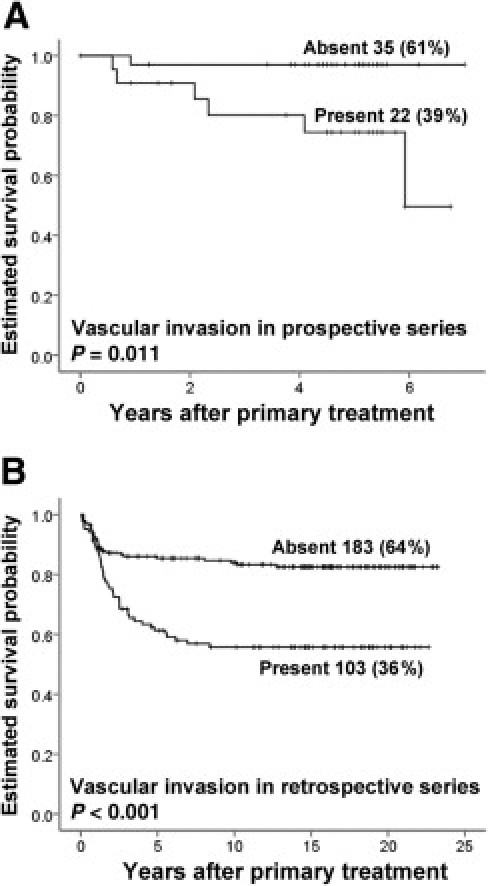
The presence of tumor cells entering vascular channels is a prognostic marker for many cancers, including endometrial carcinoma. Vascular invasion is considered to be an early step in the metastatic process and important for the progress of malignant tumors. Here, we investigated the gene expression patterns related to vascular involvement in 57 primary endometrial cancers, using DNA microarray and quantitative PCR techniques. A vascular invasion signature of 18 genes was significantly associated with patient survival and clinicopathological phenotype. Vascular involvement was also related to gene sets for epithelial-mesenchymal transition, wound response, endothelial cells, and vascular endothelial growth factor (VEGF) activity. With immunohistochemical validation, both collagen 8 and matrix metalloproteinase 3 (MMP3) were associated with vascular invasion, whereas ANGPTL4 and IL-8 were associated with patient survival. Our findings indicate that vascular involvement within primary tumors is associated with gene expression profiles related to angiogenesis and epithelial-mesenchymal transition. These data could contribute to an improved understanding of potential targets for metastatic spread and may provide clinically important information for better management of endometrial cancer.
Copyright © 2011 American Society for Investigative Pathology. Published by Elsevier Inc. All rights reserved.
Figures


Similar articles
-
Tumor necrosis is an important hallmark of aggressive endometrial cancer and associates with hypoxia, angiogenesis and inflammation responses.Oncotarget. 2015 Nov 24;6(37):39676-91. doi: 10.18632/oncotarget.5344. Oncotarget. 2015. PMID: 26485755 Free PMC article.
-
Gene alterations in tumor-associated endothelial cells from endometrial cancer.Int J Mol Med. 2008 Nov;22(5):619-32. Int J Mol Med. 2008. PMID: 18949382
-
Endometrial Carcinoma Recurrence Score (ECARS) validates to identify aggressive disease and associates with markers of epithelial-mesenchymal transition and PI3K alterations.Gynecol Oncol. 2014 Sep;134(3):599-606. doi: 10.1016/j.ygyno.2014.06.026. Epub 2014 Jul 1. Gynecol Oncol. 2014. PMID: 24995579
-
[Angiogenesis-prognostic factor in patients with endometrial cancer].Ginekol Pol. 2005 Oct;76(10):838-45. Ginekol Pol. 2005. PMID: 16417101 Review. Polish.
-
Molecular determinants of invasion in endometrial cancer.Clin Transl Oncol. 2007 May;9(5):272-7. doi: 10.1007/s12094-007-0054-z. Clin Transl Oncol. 2007. PMID: 17525037 Review.
Cited by
-
An 18-gene signature for vascular invasion is associated with aggressive features and reduced survival in breast cancer.PLoS One. 2014 Jun 6;9(6):e98787. doi: 10.1371/journal.pone.0098787. eCollection 2014. PLoS One. 2014. PMID: 24905342 Free PMC article.
-
Assessment of early response biomarkers in relation to long-term survival in patients with HER2-negative breast cancer receiving neoadjuvant chemotherapy plus bevacizumab: Results from the Phase II PROMIX trial.Int J Cancer. 2018 Feb 1;142(3):618-628. doi: 10.1002/ijc.31070. Epub 2017 Oct 13. Int J Cancer. 2018. PMID: 28940389 Free PMC article. Clinical Trial.
-
AXL modulates extracellular matrix protein expression and is essential for invasion and metastasis in endometrial cancer.Oncotarget. 2016 Nov 22;7(47):77291-77305. doi: 10.18632/oncotarget.12637. Oncotarget. 2016. PMID: 27764792 Free PMC article.
-
eIF4E‑related miR‑320a and miR‑340‑5p inhibit endometrial carcinoma cell metastatic capability by preventing TGF‑β1‑induced epithelial‑mesenchymal transition.Oncol Rep. 2020 Feb;43(2):447-460. doi: 10.3892/or.2019.7437. Epub 2019 Dec 16. Oncol Rep. 2020. PMID: 31894279 Free PMC article.
-
Establishing the Role of PPARβ/δ in Carcinogenesis.Trends Endocrinol Metab. 2015 Nov;26(11):595-607. doi: 10.1016/j.tem.2015.09.004. Epub 2015 Oct 18. Trends Endocrinol Metab. 2015. PMID: 26490384 Free PMC article. Review.
References
-
- Hanahan D., Weinberg R.A. The hallmarks of cancer. Cell. 2000;100:57–70. - PubMed
-
- Ferrari M.K., McNeal J.E., Malhotra S.M., Brooks J.D. Vascular invasion predicts recurrence after radical prostatectomy: stratification of risk based on pathologic variables. Urology. 2004;64:749–753. - PubMed
-
- Mohammed R.A., Martin S.G., Gill M.S., Green A.R., Paish E.C., Ellis I.O. Improved methods of detection of lymphovascular invasion demonstrate that it is the predominant method of vascular invasion in breast cancer and has important clinical consequences. Am J Surg Pathol. 2007;31:1825–1833. - PubMed
-
- Stefansson I.M., Salvesen H.B., Immervoll H., Akslen L.A. Prognostic impact of histological grade and vascular invasion compared with tumour cell proliferation in endometrial carcinoma of endometrioid type. Histopathology. 2004;44:472–479. - PubMed
-
- Straume O., Akslen L.A. Independent prognostic importance of vascular invasion in nodular melanomas. Cancer. 1996;78:1211–1219. - PubMed
Publication types
MeSH terms
Substances
LinkOut - more resources
Full Text Sources
Other Literature Sources
Miscellaneous
