. 2011 Apr 15;20(8):1595-609.
doi: 10.1093/hmg/ddr037. Epub 2011 Jan 31.

Mutations in PPIB (cyclophilin B) delay type I procollagen chain association and result in perinatal lethal to moderate osteogenesis imperfecta phenotypes

Affiliations

Mutations in PPIB (cyclophilin B) delay type I procollagen chain association and result in perinatal lethal to moderate osteogenesis imperfecta phenotypes

Shawna M Pyott et al. Hum Mol Genet. .

Abstract

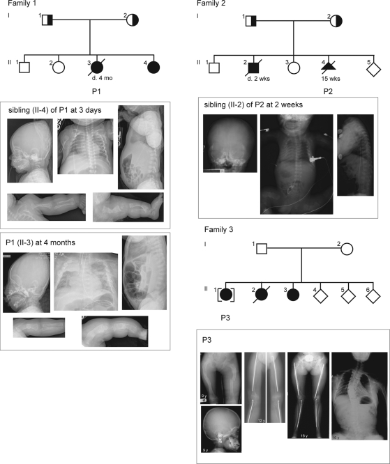
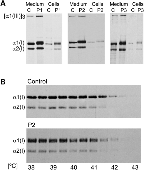
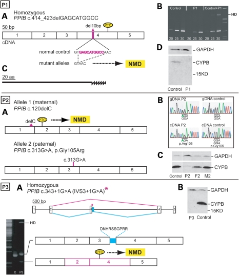
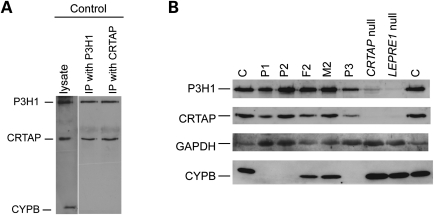
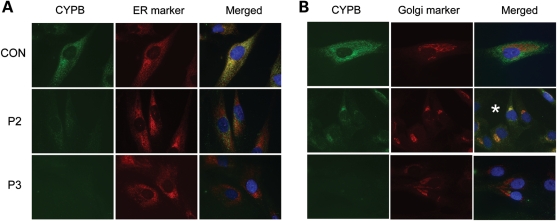
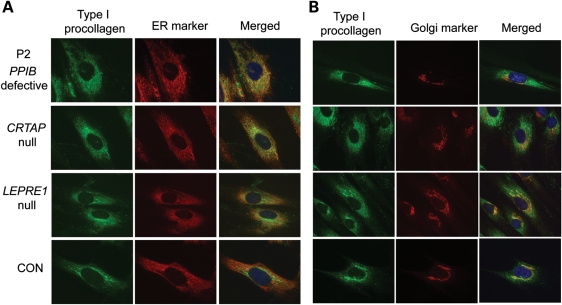
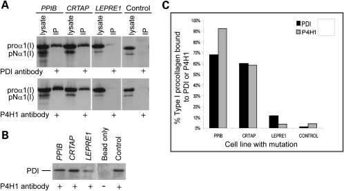
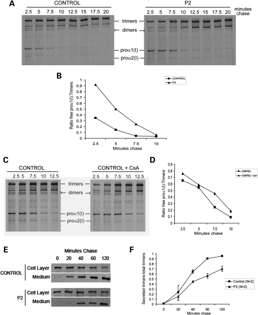
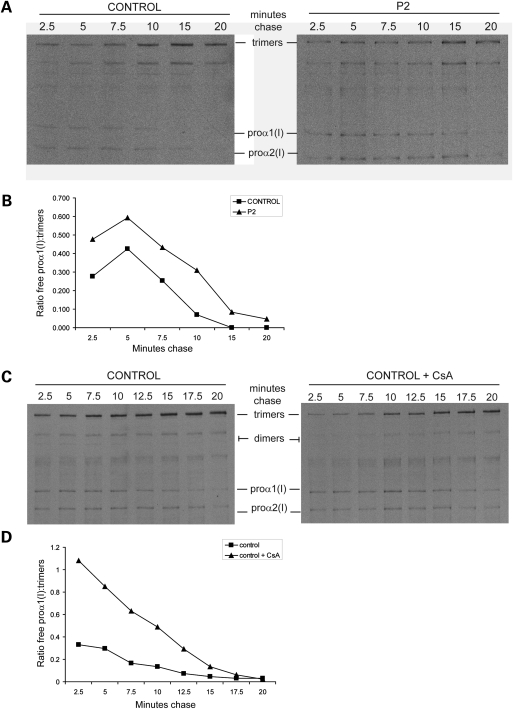
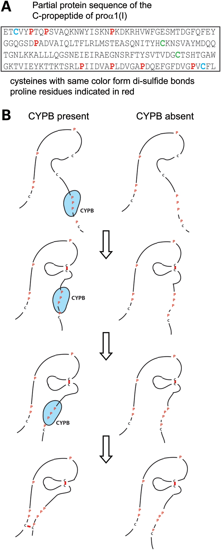
Recessive mutations in the cartilage-associated protein (CRTAP), leucine proline-enriched proteoglycan 1 (LEPRE1) and peptidyl prolyl cis-trans isomerase B (PPIB) genes result in phenotypes that range from lethal in the perinatal period to severe deforming osteogenesis imperfecta (OI). These genes encode CRTAP (encoded by CRTAP), prolyl 3-hydroxylase 1 (P3H1; encoded by LEPRE1) and cyclophilin B (CYPB; encoded by PPIB), which reside in the rough endoplasmic reticulum (RER) and can form a complex involved in prolyl 3-hydroxylation in type I procollagen. CYPB, a prolyl cis-trans isomerase, has been thought to drive the prolyl-containing peptide bonds to the trans configuration needed for triple helix formation. Here, we describe mutations in PPIB identified in cells from three individuals with OI. Cultured dermal fibroblasts from the most severely affected infant make some overmodified type I procollagen molecules. Proα1(I) chains are slow to assemble into trimers, and abnormal procollagen molecules concentrate in the RER, and bind to protein disulfide isomerase (PDI) and prolyl 4-hydroxylase 1 (P4H1). These findings suggest that although CYPB plays a role in helix formation another effect is on folding of the C-terminal propeptide and trimer formation. The extent of procollagen accumulation and PDI/P4H1 binding differs among cells with mutations in PPIB, CRTAP and LEPRE1 with the greatest amount in PPIB-deficient cells and the least in LEPRE1-deficient cells. These findings suggest that prolyl cis-trans isomerase may be required to effectively fold the proline-rich regions of the C-terminal propeptide to allow proα chain association and suggest an order of action for CRTAP, P3H1 and CYPB in procollagen biosynthesis and pathogenesis of OI.


Figures

Figure 1.
Figure 1.
Pedigrees and radiographs for individuals with autosomal recessive OI due to PPIB mutations. (Family 1) II-4 at 3 days: X-rays from the second affected child at 3 days of age show diminished calvarial mineralization; thin, beaded ribs; fractured humerus; short, bowed and undermodeled long bones of the leg. There was no overt platyspondyly. Radiographs from the first affected child (II-3) at 4 months of age demonstrated the lack of calvarial mineralization; undermineralized, broad and beaded ribs; pronounced platyspondyly; short, bowed and undermodeled long bones of the upper and lower extremities. (Family 2) Radiographs of II-2 at 2 weeks of age showed the near absence of calvarial mineralization; very short, beaded ribs; platyspondyly; undermineralized, short and bent femora. (Family 3) At 9 years of age, the femora in II-1 were broad and poorly modeled, and calvarial mineralization was near normal. By 12 years of age, there was thinning of the femoral cortex, which continued to be apparent at 16 years of age. Scoliosis had been present at early ages and was stable by age 16 years.
Figure 2.
Figure 2.
Overmodification and normal thermal stability of collagen in the presence of PPIB mutations. (A) SDS–PAGE of medium and cell layer collagens from individuals P1, P2, P3 and a control (C) showed delayed electrophoretic mobility of a population of α1(I) and α2(I) chains, following pepsin treatment of the samples to remove the propeptide extensions. (B) The thermal stability of type I collagen from P2 cells is similar to type I collagen made by controls cells.
Figure 3.
Figure 3.
Molecular basis of autosomal recessive OI due to mutations in PPIB. (P1) (A) Homozygosity for deletion of 10 bp in exon 4 (c.414_423del), shown schematically in the cDNA diagram, led to a reading frame shift and creation of a PTC 61 nt downstream in exon 4 located 41 nt from the final exon–exon boundary in mRNA. The mRNA is predicted to undergo significant NMD. (B) RT–PCR products synthesized with primers in exons 3 and 5 that yield a 343 bp product for the control, after 20, 25 and 30 cycles for a control, P1, and a 1:1 mix of the control and P1 cDNA. After 25 cycles, when the PCR was still in the linear phase, the estimated ratio of control to P1 product was 9:1. (C) The remaining small amount of mutant mRNA in P1 would result in a shortened protein of 158 amino acids (full length is 216 amino acids) with the last 20 amino acids being different from normal (serrated part of the line diagram). (D) Western blot analysis using a polyclonal antibody directed against a full-length recombinant protein of human CYPB did not detect the predicted shortened fragment or a full-length fragment (see Materials and methods). (P2) (A) Compound heterozygosity for c.120delC (maternal allele) and c.313G > A, p.Gly105Arg (paternal allele). The single-nucleotide deletion resulted in a reading frame shift and a PTC 49 nt downstream in exon 2. (B) In genomic DNA (gDNA), P2 is heterozygous A/G at c.313, whereas in cDNA only the A allele is present because mRNA from the G allele that harbors the c.120delC mutation is rapidly degraded. (C) Western blot using a polyclonal antibody directed against a region within residues 150 to the C-terminus of human CYPB showed only a very small amount of CYPB protein derived from the stable mRNA in P2, whereas the amount in the cells from the carrier parents (F2-father, M2-mother) appeared to be close to normal. (P3) (A) Homozygosity for an intron 3 splice donor site mutation (c.343 + 1G > A, IVS3 + 1G > A) yielded two products from each allele. In the first, 27 nucleotides of intron 3 were retained in the mature mRNA due to the use of a strong cryptic splice donor site starting at IVS3 + 28 (gtatgt), which resulted in removal of one glycine residue (p.Gly115) and insertion of 10 new amino acids that are shown in single letter code: DNHRSSGPRR. In the second, exon 3 (94 nt) was skipped, which resulted in a reading frame shift and a PTC in exon 4 that was predicted to lead to NMD (HD, heteroduplex). (B) Western blot analysis using a polyclonal antibody directed against a synthetic peptide corresponding to residues 194–208 of human CYPB did not detect any CYPB protein.
Figure 4.
Figure 4.
Mutations in PPIB and loss of CYPB do not destabilize CRTAP or P3H1. (A) Analysis of P3H1, CRTAP and CYPB protein interactions in control cells. The proteins in the entire cell lysate or proteins brought down with antibody to P3H1or CRTAP were separated by SDS–PAGE on 10% polyacrylamide gels and then stained with a pool of antibodies to P3H1 (from Kevin McCarthy), CRTAP (from Roy Morello, see Materials and methods) and CYPB (Abcam). Although CYPB is present in the lysate, it is not brought down at a detectable level by antibodies to the two other proteins. (B) Effects of mutations in PPIB, CRTAP and LEPRE1 on stability of other proteins in the complex. In cells with mutations in PPIB that resulted in loss (P1 and P3) or reduction (P2) in the CYPB protein (antibodies used for CYPB analysis for each proband are indicated in the Results section), the stabilities of CRTAP and P3H1 were unaffected. Parents of P2 (M2 and F2) showed a reduced amount of CYPB protein as expected. In cells with mutations in either CRTAP or LEPRE1, there is a marked reduction or complete loss of the normal protein partner, whereas levels of CYPB remained unaffected. GAPDH was used as a loading control.
Figure 5.
Figure 5.
Abnormal CYPB protein in P2 fibroblasts mis-localizes to the Golgi. (A) Immunocytochemistry with antibodies against CYPB and an RER marker showed CYPB protein localized to the RER in control fibroblasts, reduction in CYPB in P2 fibroblasts (antibody used for P2 listed in the Results section) and loss of CYPB in P3 fibroblasts (antibody used for P3 listed in the Results section). (B) Staining with CYPB and a Golgi marker showed the abnormal CYPB protein produced by P2 fibroblasts mis-localized to the Golgi (indicated by a white asterisk). In the P3 fibroblasts, CYPB was not evident.
Figure 6.
Figure 6.
Retention of type I procollagen in the RER differs among cells with mutations in PPIB, CRTAP and LEPRE1 (A and B). Type I procollagen is selectively retained in the RER in cells with mutations in PPIB, whereas cells with mutations in CRTAP or LEPRE1 distribute the molecules between the RER and the Golgi. In contrast, the majority of type I procollagen is located in the Golgi in control cells.
Figure 7.
Figure 7.
Type I procollagen binds to PDI and P4H1 in the RER. (A) Proteins in lysates of cells with mutations in PPIB (P2), CRTAP or LEPRE1 were precipitated with antibodies to PDI or P4H1 and then stained with antibodies to proα1(I) (LF9). The greatest proportion of available proα1(I) chains bound by PDI and P4H1 was seen in the PPIB mutant cells with decreasing amounts in CRTAP, LEPRE1 mutant cells and the least bound in control cells, quantitated in (C). (B) PDI precipitated by antibody to P4H1 in cells from individuals with mutations indicated in (A).
Figure 8.
Figure 8.
Proα1(I) chains in P2 fibroblasts and control cells treated with CsA are slow to assemble and secretion of type I procollagen is delayed. (A and C) Cells were labeled for 10 min with [35S]-methionine and cysteine, immunoprecipitated with the LF9 antibody directed to the N-terminal propeptide of proα1(I) chains, and then analyzed by SDS–PAGE under non-reducing conditions. (B and D) P2 fibroblasts and control fibroblasts treated with 5 µm CsA had more free proα1(I) for up to 10 min compared with controls, and trimers accumulated in the cell over a longer period. (E) Cells were labeled with [3H]-proline for 60 min, chased for up to 2 h with proteins harvested every 20 min. The protein lysates were analyzed by SDS–PAGE under non-reducing conditions. (F) The relative amount of type I procollagen trimers in the medium was far greater in control cells than in the P2 cells. Only the trimers are shown in all four gels.
Figure 9.
Figure 9.
Proα1(I) chains in CsA-treated control fibroblasts and P2 fibroblasts are slow to assemble. (A) Control and P2 fibroblasts were labeled for 15 min with [3H]-proline, the cells lysed and the proteins analyzed by SDS–PAGE under non-reducing conditions. Density of the protein bands were quantitated using ImageJ software. (B) P2 fibroblasts had more free proα1(I) at each time point compared with control fibroblasts, and P2 trimers were slow to assemble compared with trimers in control cells. (C) Control fibroblasts were labeled the same as in (A) then treated with 5 µm CsA or EtOH alone. (D) Fibroblasts treated with 5 µm CsA had more free proα1(I) at each time point compared with control fibroblasts with no drug treatment, and trimers were slow to form in the drug-treated cells compared with control cells.
Figure 10.
Figure 10.
Schematic of proposed model. (A) The sequence of the C-terminal propeptide of proα1(I) chains with interacting cysteine residues and prolyl residues indicated. (B) The rate at which intra-chain disulfide bonds form in the C-terminal propeptide is determined by the folding of the proline-rich region around cysteine residues, a region that is likely a substrate for CYPB.

References

    1. Morello R., Bertin T., Chen Y., Hicks J., Tonachini L., Monticone M., Castagnola P., Rauch F., Glorieux F., Vranka J., et al. CRTAP is required for prolyl 3- hydroxylation and mutations cause recessive osteogenesis imperfecta. Cell. 2006;127:291–304. doi:10.1016/j.cell.2006.08.039. - DOI - PubMed
    1. Barnes A., Chang W., Morello R., Cabral W., Weis M., Eyre D., Leikin S., Makareeva E., Kuznetsova N., Uveges T., et al. Deficiency of cartilage-associated protein in recessive lethal osteogenesis imperfecta. N. Engl. J. Med. 2006;355:2757–2764. doi:10.1056/NEJMoa063804. - DOI - PMC - PubMed
    1. Cabral W., Chang W., Barnes A., Weis M., Scott M., Leikin S., Makareeva E., Kuznetsova N., Rosenbaum K., Tifft C., et al. Prolyl 3-hydroxylase 1 deficiency causes a recessive metabolic bone disorder resembling lethal/severe osteogenesis imperfecta. Nat. Genet. 2007;39:359–365. doi:10.1038/ng1968. - DOI - PMC - PubMed
    1. Baldridge D., Schwarze U., Morello R., Lennington J., Bertin T., Pace J., Pepin M., Weis M., Eyre D., Walsh J., et al. CRTAP and LEPRE1 mutations in recessive osteogenesis imperfecta. Hum. Mutat. 2008;29:1435–1442. doi:10.1002/humu.20799. - DOI - PMC - PubMed
    1. Willaert A., Malfait F., Symoens S., Gevaert K., Kayserili H., Megarbane A., Mortier G., Leroy J., Coucke P., De Paepe A. Recessive osteogenesis imperfecta caused by LEPRE1 mutations: clinical documentation and identification of the splice form responsible for prolyl 3-hydroxylation. J. Med. Genet. 2009;46:233–241. doi:10.1136/jmg.2008.062729. - DOI - PubMed

Publication types

MeSH terms