. 2011 Apr 1;589(Pt 7):1603-18.
doi: 10.1113/jphysiol.2010.201939. Epub 2011 Jan 31.

The role of synaptobrevin1/VAMP1 in Ca2+-triggered neurotransmitter release at the mouse neuromuscular junction

Affiliations

The role of synaptobrevin1/VAMP1 in Ca2+-triggered neurotransmitter release at the mouse neuromuscular junction

Yun Liu et al. J Physiol. .

Abstract

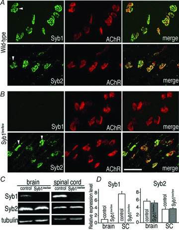
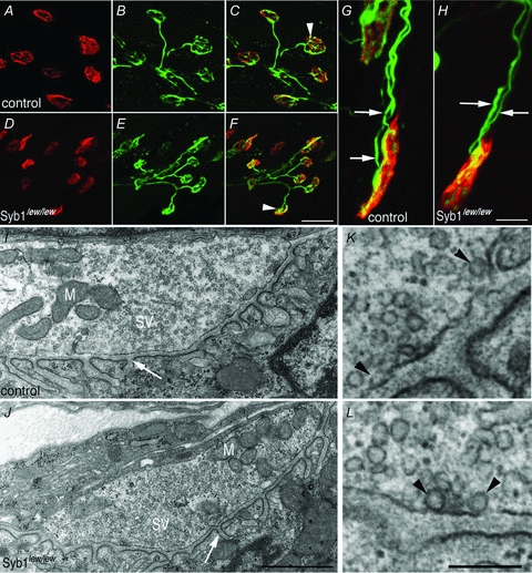
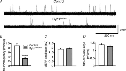
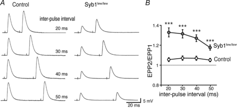
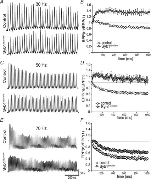
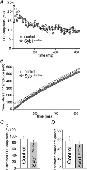
Synaptobrevin (Syb)/vesicle-associated membrane protein (VAMP) is a small, integral membrane protein of synaptic vesicles. Two homologous isoforms of synaptobrevin, Syb1/VAMP1 and Syb2/VAMP2, exhibit distinct but partially overlapping patterns of expression in adult mammalian neurons: Syb1 is predominantly expressed in the spinal cord, especially in motor neurons and motor nerve terminals of the neuromuscular junction (NMJ), whereas Syb2 is primarily expressed in central synapses in the brain. Whereas many studies have focused on the function of Syb2 in the brain, few studies have examined the role of Syb1. Here we report that Syb1 plays a critical role in neuromuscular synaptic transmission. A null mutation of Syb1 resulting from a spontaneous, nonsense mutation in mice significantly impairs the function, but not the structure, of the NMJ. In particular, both spontaneous and evoked synaptic activities in Syb1 mutant mice are reduced significantly relative to control mice. Short-term synaptic plasticity in Syb1-deficient NMJs is markedly altered: paired-pulse facilitation is significantly enhanced, suggesting a reduction in the initial release probability of synaptic vesicles. Furthermore, Syb1-deficient NMJs display a pronounced asynchrony in neurotransmitter release. These impairments are not due to an alteration of the size of the readily releasable pool of vesicles, but are attributable to reduced sensitivity and cooperativity to calcium (Ca2+) due to the absence of Syb1. Our findings demonstrate that Syb1 plays an essential, non-redundant role in Ca2+-triggered vesicle exocytosis at the mouse NMJ.


Figures

Figure 1
Figure 1. Expression of Syb1 and Syb2 in WT and Syb1lew/lew mice
A and B, whole mounts of triangularis sterni (P14) muscles from WT mice (A) or Syb1lew/lew mutant mice (B) were immunostained with antibodies against Syb1 or Syb2, as well as Texas Red-conjugated α-bgt, which labels postsynaptic AChRs. Both Syb1 and Syb2 were detected at the WT nerve terminals (arrowheads in A). Syb1 was absent from Syb1lew/lew NMJs: only α-bgt staining was detected in the synaptic regions of the muscle (upper row in B). In contrast, Syb2 was detected in the nerve terminals (arrowheads in B) of Syb1lew/lew mutant mice. C, images of quantitative Western blot analysis of the brain and spinal cord (SC) from Syb1lew/lew and their littermate control mice (P14). D, comparison of relative levels of Syb1 or Syb2 between Syb1lew/lew mice (n = 3) and their littermates (control, n = 3). Relative expression levels of Syb1 and Syb2 were calculated by normalizing to that of α-tubulin (loading control). Syb1 was absent from Syb1lew/lew mice, but present in control mice, with significantly higher (P < 0.001) expression in the SC (7.73 ± 0.66, n = 3) compared to the brain (0.96 ± 0.21, n = 3) (normalized to α-tubulin level). In both Syb1lew/lew and control mice, Syb2 was more abundantly expressed in the brain compared to the SC (control: brain, 5.69 ± 0.57; SC, 3.54 ± 0.5, P < 0.05; Syb1lew/lew mutant: brain, 5.22 ± 0.57, SC, 3.69 ± 0.27, P < 0.05). In both the brain and SC, the relative levels of Syb2 remained similar between the Syb1lew/lew and control mice, P > 0.05. Scale bar in A and B, 50 μm.
Figure 2
Figure 2. Formation of neuromuscular synapses in Syb1lew/lew mice
A–H, diaphragm muscles (P14) of control (A–C and G) and Syb1lew/lew mice (D–F and H) were doubly labelled with presynaptic markers (anti-synaptotagmin 2 and anti-neurofilament) (green) and postsynaptic markers (Texas Red-conjugated α-bgt) (red). In both control and Syb1lew/lew mice, nerve terminals (B and E) formed close apposition with postsynaptic AChRs (A and D), as shown in the merged images (white arrowheads in C and F). The majority (99%) of the end-plates were innervated by a single axon, except a small number (1%) of end-plates which were occupied by two axons in both control (G, white arrows point to two individual axons) and Syb1lew/lew mice (H, white arrows). I–L, electron micrographs of longitudinal sections of P14 diaphragm muscles. A typical nerve terminal from control (I) and Syb1lew/lew mice (J): both nerve terminals were packed with abundant synaptic vesicles (SV) and mitochondria (M). Postsynaptic junctional folds (white arrow in I and J) were morphologically normal in Syb1lew/lew mice compared with the control. K and L, examples of docked vesicles (black arrowheads) viewed under high magnification. K: control; L: Syb1lew/lew. Scale bars: A–F, 20 μm; G and H, 5 μm; I and J, 1 μm; K and L, 0.2 μm.
Figure 3
Figure 3. Reduction of spontaneous neurotransmitter release at the NMJ in Syb1lew/lew mice
A, representative mEPP traces recorded from the diaphragm muscles (P14). Each trace represents a total of 2 min continuous recording from the same NMJ and is displayed as 120 superimposed 1 s sweeps. mEPP frequencies were markedly reduced in Syb1lew/lew mice compared with controls. B–D, quantitative analyses of mEPP frequencies, amplitudes and 10–90% rise slopes. The average mEPP frequencies were reduced to 3.2 ± 0.4 events min−1 in Syb1lew/lew mice, significantly lower than those in the controls (8.9 ± 1.1 events min−1) (B). mEPP amplitudes (C) of Syb1lew/lew mice (1.92 ± 0.09 mV) were similar to those of the controls (1.86 ± 0.08 mV). The 10–90% rise slopes (D) of Syb1lew/lew mice (1.3 ± 0.1 mV ms−1) and control mice (1.4 ± 0.1 mV ms−1) also were similar. Syb1lew/lew, n = 34 muscle fibres; control, n = 40 muscle fibres. ***P < 0.001, Student's t test.
Figure 4
Figure 4. Reduction of evoked neurotransmitter release at the NMJ in Syb1lew/lew mice
A, 10 consecutive EPP traces recorded from P14 diaphragm muscles (evoked by 0.2 Hz stimulation of the phrenic nerves) are superimposed. EPPs in the control mice were highly reproducible; the peaks of all 10 EPPs, as indicated by circles located to left of the traces, all fall within a range of 7 mV (between 20 and 27 mV). In contrast, EPPs in Syb1lew/lew mice were highly variable, fluctuating over a range of 15 mV (between 2 and 17 mV, filled circles). An F test demonstrated that the variances in Syb1lew/lew mice were significantly (*P < 0.05) increased compared to those of the controls. Similarly, EPP rising phases (arrowhead) and decay phases (arrow) were also highly variable in Syb1lew/lew mice compared with control mice. B, quantification of EPP amplitudes between control and Syb1lew/lew mice: EPP amplitudes were significantly reduced in Syb1lew/lew mice (15.5 ± 1.2 mV), compared with those in the control mice (25.9 ± 1.3 mV). C, the EPP rise-slopes (10–90%) in Syb1lew/lew mice (10.3 ± 0.8 mV ms−1) were significantly reduced compared to those in the controls (17.3 ± 0.9 mV ms−1). D, quantal contents were significantly reduced in Syb1lew/lew mice (8.6 ± 0.7) compared to controls (16.1 ± 0.6). Syb1lew/lew, n = 25 muscle fibres; control, n = 22 muscle fibres. ***P < 0.001, Student's t test.
Figure 5
Figure 5. Impairment of paired-pulse facilitation at the NMJ in Syb1lew/lew mice
A, sample EPP traces recorded from P14 diaphragm muscles following twin-pulse stimulation of the phrenic nerves at various interpulse intervals (20–50 ms). B, paired pulse ratio (EPP2/EPP1) plotted as a function of the inter-pulse-interval. Syb1lew/lew NMJs exhibited enhanced facilitation compared with control NMJs. Number of NMJs: n = 21, Syb1lew/lew; n = 18, control. ***P < 0.001, Student's t test.
Figure 6
Figure 6. Altered short-term neuromuscular synaptic plasticity in Syb1lew/lew mice
Left (A, C and E), representative EPP traces responded to 30 Hz (A), 50 Hz (C) or 70 Hz (E) train stimulation (1 s train). Right (B, D and F), EPP run-down calculated by the ratio of EPP amplitude to the first EPP amplitude during 30, 50 and 70 Hz stimulation. In the control NMJ, EPP ratios were progressively decreased during the course of train stimulation at 30, 50, and 70 Hz, with more pronounced depression at higher frequency (70 Hz > 50 Hz > 30 Hz). In contrast, EPP amplitudes in Syb1lew/lew NMJs were facilitated during 30 Hz and 50 Hz stimulation, as well as during the first few traces of 70 Hz stimulation (F). At all three frequencies (30, 50 and 70 Hz), the rate of EPP run-down in Syb1lew/lew NMJs (n = 19) was significantly reduced compared to the control (n = 14) (P < 0.001, Student's t test).
Figure 7
Figure 7. Normal size of readily releasable pool (RRP) at the NMJ in Syb1lew/lew mice
A, analyses of EPP amplitudes in the diaphragm muscle (P14) evoked by a high frequency repetitive stimulation (70 Hz) of the phrenic nerve. The peak of EPP amplitudes reaches a steady state after 40–44 stimuli in both control and Syb1lew/lew NMJs. The first 44 EPPs are shown in this plot. B, cumulative EPP amplitudes as shown in A. Five data points from 40th to 44th stimulus were fitted by linear regression and back-extrapolated to time zero (control: black dashed line; Syb1lew/lew: grey dashed line). C, estimated EPP amplitudes at time zero. D, estimated number of quanta obtained by dividing the estimated EPP amplitudes at time zero in C with mEPP amplitude of the same cell. The resulting estimated number of quanta is similar between the control (57.2 ± 8.9, n = 8) and Syb1lew/lew (50.4 ± 5.9, n = 9; P > 0.05), suggesting the size of the RRP is normal in Syb1lew/lew NMJs.
Figure 8
Figure 8. Reduced Ca2+ sensitivity and cooperativity of neuromuscular synaptic transmission in Syb1lew/lew mice
A, sample EPP traces recorded at various external [Ca2+] (0.5, 1.0, 2.0 and 4.0 mm). Each panel represents 10 superimposed EPP traces, with peaks indicated by open circles on the left side. In both control (A) and Syb1lew/lew mice (B), EPP amplitudes decrease and variability increases as external [Ca2+] is reduced. C and D, dose–response curve of average EPP amplitude (C) and quantal content (D), as a function of external [Ca2+], from recordings of P14 diaphragm muscles from Syb1lew/lew and control mice. The curve is fitted by a sigmoidal dose–response equation: formula image
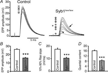
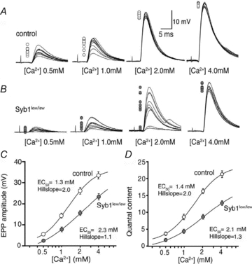
, where Y is the value of the EPP amplitude or the quantal content at the indicated [Ca2+], Ymax and Ymin are the maximum and minimum EPP amplitude or quantal content, respectively, EC50 is the [Ca2+] when Y is 50% of Ymax, and k is the Hill coefficient (Hill slope), which reflects the steepness of the curve. The sigmoidal dose–response curve of EPP amplitude and quantal content as a function of external [Ca2+] showed that the EC50 was markedly increased and the Hill slope was markedly reduced in Syb1lew/lew NMJs (n = 29) compared with the control NMJs (n = 23).
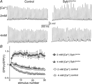
Figure 9
Figure 9. Elevating external [Ca2+] restores short-term plasticity of Syb1lew/lew NMJs
A, sample EPP traces of control and Syb1lew/lew NMJs (P14 diaphragm muscle) evoked by 30 Hz, 1 s train stimulation of the phrenic nerve at 2 mm[Ca2+] and 4 mm[Ca2+]. B, quantification of the EPP amplitudes shown in A. EPP amplitudes were facilitated during 30 Hz stimulation in Syb1lew/lew NMJs (n = 29) at 2 mm[Ca2+] (filled circles), and this facilitation was eliminated when [Ca2+] was increased to 4 mm (filled triangles), approaching the normal EPP run-down typically exhibited in control NMJs (n = 24) at 2 mm[Ca2+] (open circles).

References

    1. Arac D, Chen X, Khant HA, Ubach J, Ludtke SJ, Kikkawa M, Johnson AE, Chiu W, Sudhof TC, Rizo J. Close membrane-membrane proximity induced by Ca2+-dependent multivalent binding of synaptotagmin-1 to phospholipids. Nat Struct Mol Biol. 2006;13:209–217. - PubMed
    1. Augustine GJ, Charlton MP, Smith SJ. Calcium action in synaptic transmitter release. Annu Rev Neurosci. 1987;10:633–693. - PubMed
    1. Balice-Gordon RJ, Lichtman JW. In vivo observations of pre- and postsynaptic changes during the transition from multiple to single innervation at developing neuromuscular junctions. J Neurosci. 1993;13:834–855. - PMC - PubMed
    1. Baumert M, Maycox PR, Navone F, De Camilli P, Jahn R. Synaptobrevin: an integral membrane protein of 18,000 daltons present in small synaptic vesicles of rat brain. EMBO J. 1989;8:379–384. - PMC - PubMed
    1. Bennett MK, Calakos N, Scheller RH. Syntaxin: a synaptic protein implicated in docking of synaptic vesicles at presynaptic active zones. Science. 1992;257:255–259. - PubMed

Publication types

MeSH terms

Substances