. 2011 Feb 14;208(2):261-71.
doi: 10.1084/jem.20101688. Epub 2011 Jan 31.

Bone marrow CD169+ macrophages promote the retention of hematopoietic stem and progenitor cells in the mesenchymal stem cell niche

Affiliations

Bone marrow CD169+ macrophages promote the retention of hematopoietic stem and progenitor cells in the mesenchymal stem cell niche

Andrew Chow et al. J Exp Med. .

Abstract

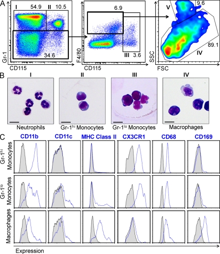
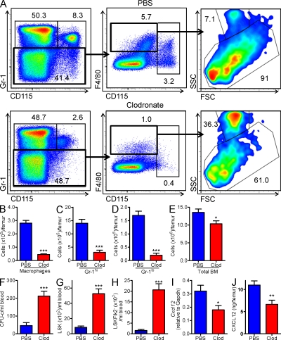
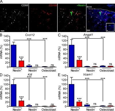
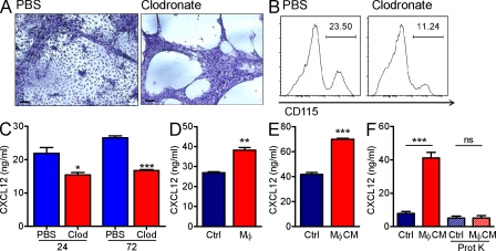
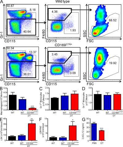
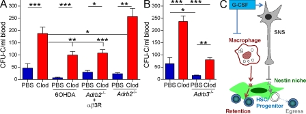
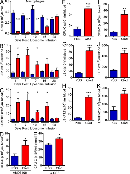
Hematopoietic stem cells (HSCs) reside in specialized bone marrow (BM) niches regulated by the sympathetic nervous system (SNS). Here, we have examined whether mononuclear phagocytes modulate the HSC niche. We defined three populations of BM mononuclear phagocytes that include Gr-1(hi) monocytes (MOs), Gr-1(lo) MOs, and macrophages (MΦ) based on differential expression of Gr-1, CD115, F4/80, and CD169. Using MO and MΦ conditional depletion models, we found that reductions in BM mononuclear phagocytes led to reduced BM CXCL12 levels, the selective down-regulation of HSC retention genes in Nestin(+) niche cells, and egress of HSCs/progenitors to the bloodstream. Furthermore, specific depletion of CD169(+) MΦ, which spares BM MOs, was sufficient to induce HSC/progenitor egress. MΦ depletion also enhanced mobilization induced by a CXCR4 antagonist or granulocyte colony-stimulating factor. These results highlight two antagonistic, tightly balanced pathways that regulate maintenance of HSCs/progenitors in the niche during homeostasis, in which MΦ cross talk with the Nestin(+) niche cell promotes retention, and in contrast, SNS signals enhance egress. Thus, strategies that target BM MΦ hold the potential to augment stem cell yields in patients that mobilize HSCs/progenitors poorly.


Figures

Figure 1.
Figure 1.
Mononuclear phagocytes can be distinguished in the BM with Gr-1, CD115, and F4/80. (A) Gating strategy of BM mononuclear phagocytes. The Gr-1+ populations were divided into a CD115 fraction comprised of neutrophils (I; left) and CD115+ fraction of Gr-1hi MOs (II; left). The Gr-1lo fraction was further subdivided into two populations (middle): CD115+ Gr-1lo MOs (III) and a F4/80+CD115int population, which can be subdivided into SSChi eosinophils (V) and SSCint/lo MΦ (IV). (B) Morphology of cell populations I–IV (63× magnification; bars, 10 µm). (C) Overlay histograms show the differential expression of CD11b, CD11c, MHC class II, CX3CR1, CD68, and CD169 (blue line) among mononuclear phagocyte populations. Gray histograms represent isotype control. All results are representative of two independent experiments.
Figure 2.
Figure 2.
Depletion of mononuclear phagocytes is associated with HSC/progenitor mobilization and CXCL12 reduction. (A–H) C57BL/6 mice were treated with PBS (blue) or clodronate-encapsulated liposomes (red). (A) Representative dot plots show the percentages of neutrophils, Gr-1hi MOs, Gr-1lo MOs, and MΦ, as described in Fig. 1 A. (B–E) Absolute numbers of mononuclear phagocytes and total nucleated cells in the BM (n = 11). (F) Absolute numbers of colony-forming units in culture in the peripheral blood (CFU-C; n = 12–15). (G–H) Enumeration of Lineage Sca-1+ c-kit+ (LSK; G) and LSKFlk2 (H) cells in the peripheral blood (n = 9–10). B–H represents pooled data from at least three independent experiments. (I) RT-PCR analysis of Cxcl12 mRNA levels in total BM cells (n = 5 mice per group). Data representative of two independent experiments are shown. (J) CXCL12 levels in the BMEF. Data are pooled from three independent experiments.
Figure 3.
Figure 3.
Anatomical and functional relationships between mononuclear phagocytes and Nestin+ niche cells. (A) Distribution of BM CD68+ (white) and CD169+ (red) cells in 5-µm femoral sections of Nes-GFP mice. Dashed line demarcates separation of bone and BM. Images were acquired at 40× magnification (bar, 20 µm). Isotype control is shown in inset. (B–E) Relative expression of Cxcl12 (B), Angpt1 (C), Kitl (D), and Vcam1 (E) in CD45 Ter119 Nestin+ (Nestin+) and CD45 Ter119 Nestin (Nestin) fractions sorted from the BM and osteoblasts sorted from the bone 14 h after treatment with PBS- (blue bars) or clodronate-encapsulated (red bars) liposomes. Data are presented with the mean expression of the PBS liposome-treated Nestin+ fraction set at 100% and are representative of three independent experiments (n = 4–5). Data analyzed by one-way ANOVA/Newman-Keuls test. ***, P < 0.001.
Figure 4.
Figure 4.
MΦ in culture promote CXCL12 production. (A) Morphology of adherent layer of Dexter culture 24 h after addition of PBS or clodronate liposomes (10×; bars, 100 µm). (B) Adherent cells were analyzed by flow cytometry for CD115+ cells. (A and B) Representative data from two independent experiments are shown. (C) CXCL12 levels were assessed by ELISA at 24 h (left bars) and 72 h (right bars) after liposomal incubation. (D) CXCL12 levels were assessed 3 d after MS-5 cells were co-cultured with (MΦ) or without (Ctrl) BMDM. (E) Levels of CXCL12 secreted from MS-5 cells after culture with medium conditioned by BMDM (MΦ CM) or with control medium (Ctrl). (F) CXCL12 levels were measured after culture of MS-5 cells with control (Ctrl, blue) or MΦ-conditioned (MΦ CM, red) medium that was untreated (left two bars) or treated with Proteinase K (right two bars). (C–F) Representative data from at least two independent experiments are shown.
Figure 5.
Figure 5.
Depletion of BM CD169+ MΦ, but not CD169 MOs, mobilizes HSCs/progenitors. (A–D) Wild-type (WT) or heterozygous CD169-DTR (CD169DTR/+) mice were treated with DT. (A) Representative dot plots show the percentages of BM mononuclear phagocytes in wild-type (top) or CD169DTR/+ mice treated with DT (bottom). (B–D) Bar graphs depict the absolute numbers of mononuclear phagocytes (n = 6) in wild type mice and wild type or CD169DTR/+ mice injected with DT. (E–F) Bar graphs enumerate Lineage Sca-1+ c-kit+ (LSK; E) and LSKFlk2cells (F) per milliliter of blood in the same mice analyzed in B–D (n = 6). Data are pooled from two independent experiments. (G) CXCL12 levels were measured 72 h after administration of PBS or 1 µg/ml DT into Dexter cultures plated from the BM of CD169-DTR animals (n = 3–4 wells). Representative data from two independent experiments are shown.
Figure 6.
Figure 6.
Opposite influences of the MΦ and the SNS on HSC/progenitor retention. (A) CFU-C after treatment with PBS (blue bars) or clodronate liposomes (red bars) in wild-type C57BL/6 (n = 11–13), 6OHDA-treated mice (n = 12–13), β2-adrenergic receptor–deficient (Adrb2−/−) mice treated with an antagonist to the β3-adrenergic receptor (αβ3R; n = 10), and Adrb2−/− mice (n =7). Data are pooled from three independent experiments and analyzed with one-way ANOVA/Newman-Keuls test. (B) CFU-C after treatment with PBS (blue bars) or clodronate (red bars) in wild-type FVB mice (n = 5) and β3-adrenergic receptor-deficient (Adrb3−/−) mice (n = 8). Data are pooled from two independent experiments. (C) Schematic of antagonistic regulation of HSC/progenitor retention by MΦ and the SNS.
Figure 7.
Figure 7.
MΦ depletion synergizes with AMD3100 and G-CSF mobilization. (A–C) Kinetics of MΦ reduction (A) and LSK (B) and LSKFlk2 (C) mobilization, at the indicated time points, after administration of PBS- (blue) or clodronate-encapsulated (red) liposomes (n = 3–4). Data are pooled from two independent experiments. (D–E) CFU-C from peripheral blood of mice that were treated with PBS (blue) or clodronate liposomes (red; 14 h before harvest) and mobilized with AMD3100 (D; 1 h before harvest) or G-CSF (E; 4 d). Data are pooled from two independent experiments. (F–H) CFU-C (F), LSK (G), and LSKFlk2 (H) cells from the peripheral blood of mice that were mobilized for 2 d with G-CSF and treated with PBS- or clodronate-encapsulated liposomes 14 h before harvest. Experiment was performed once (n = 4). (I–K) CFU-C (I), LSK (J), and LSKFlk2 (K) from 16-wk-old female mice that were pretreated with PBS- or clodronate-encapsulated liposomes 10 d before harvest and mobilized with G-CSF for 4 d. Data are representative of two independent experiments (n = 4).

References

    1. Balordi F., Fishell G. 2007. Mosaic removal of hedgehog signaling in the adult SVZ reveals that the residual wild-type stem cells have a limited capacity for self-renewal. J. Neurosci. 27:14248–14259 10.1523/JNEUROSCI.4531-07.2007 - DOI - PMC - PubMed
    1. Bensinger W., DiPersio J.F., McCarty J.M. 2009. Improving stem cell mobilization strategies: future directions. Bone Marrow Transplant. 43:181–195 10.1038/bmt.2008.410 - DOI - PubMed
    1. Berg D.J., Davidson N., Kuhn R., Muller W., Menon S., Holland G., Thompson-Snipes L., Leach M.W., Rennick D. 1996. Enterocolitis and colon cancer in interleukin-10-deficient mice are associated with aberrant cytokine production and CD4(+) TH1-like responses. J. Clin. Invest. 98:1010–1020 10.1172/JCI118861 - DOI - PMC - PubMed
    1. Buch T., Heppner F.L., Tertilt C., Heinen T.J., Kremer M., Wunderlich F.T., Jung S., Waisman A. 2005. A Cre-inducible diphtheria toxin receptor mediates cell lineage ablation after toxin administration. Nat. Methods. 2:419–426 10.1038/nmeth762 - DOI - PubMed
    1. Burnett S.H., Kershen E.J., Zhang J., Zeng L., Straley S.C., Kaplan A.M., Cohen D.A. 2004. Conditional macrophage ablation in transgenic mice expressing a Fas-based suicide gene. J. Leukoc. Biol. 75:612–623 10.1189/jlb.0903442 - DOI - PubMed

Publication types

MeSH terms