

Deciphering the mode of action of the synthetic antimicrobial peptide Bac8c
- PMID: 21282431
- PMCID: PMC3067151
- DOI: 10.1128/AAC.01053-10
Deciphering the mode of action of the synthetic antimicrobial peptide Bac8c
Abstract
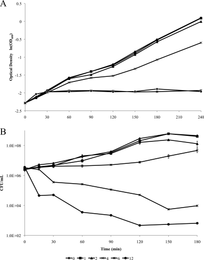
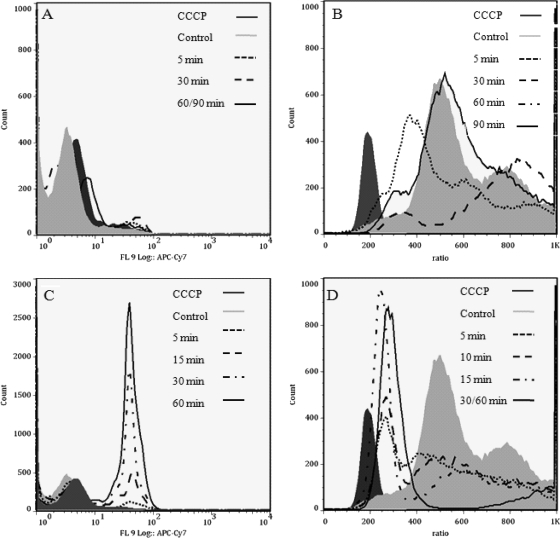
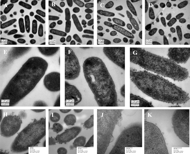
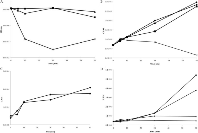
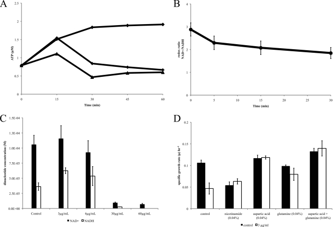
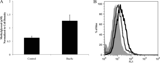
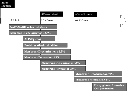
Bac8c (RIWVIWRR-NH(2)) is an 8-amino-acid peptide derived from Bac2A (RLARIVVIRVAR-NH(2)), a C3A/C11A variant of the naturally occurring bovine peptide, bactenecin (also known as bovine dodecapeptide), the smallest peptide with activity against a range of pathogenic Gram-positive and Gram-negative bacteria, as well as yeast. The effects of Bac8c on Escherichia coli were examined by studying its bacteriostatic and bactericidal properties, demonstrating its effects on proton motive force generation, and visually analyzing (via transmission electron microscopy) its effects on cells at different concentrations, in order to probe the complexities of the mechanism of action of Bac8c. Results were consistent with a two-stage model for the Bac8c mode of action. At sublethal concentrations (3 μg/ml), Bac8c addition resulted in transient membrane destabilization and metabolic imbalances, which appeared to be linked to inhibition of respiratory function. Although sublethal concentrations resulted in deleterious downstream events, such as methylglyoxal formation and free radical generation, native E. coli defense systems were sufficient for full recovery within 2 h. In contrast, at the minimal bactericidal concentration (6 μg/ml), Bac8c substantially but incompletely depolarized the cytoplasmic membrane within 5 min and disrupted electron transport, which in turn resulted in partial membrane permeabilization and cell death.
Figures
Similar articles
-
Lipoic acid modified antimicrobial peptide with enhanced antimicrobial properties.Bioorg Med Chem. 2020 Oct 1;28(19):115682. doi: 10.1016/j.bmc.2020.115682. Epub 2020 Aug 2. Bioorg Med Chem. 2020. PMID: 32912428
-
Fungicide Bac8c triggers attenuation of mitochondrial homeostasis and caspase-dependent apoptotic death.Biochimie. 2017 Feb;133:80-86. doi: 10.1016/j.biochi.2016.12.013. Epub 2016 Dec 25. Biochimie. 2017. PMID: 28027901
-
Fungicidal mechanisms of the antimicrobial peptide Bac8c.Biochim Biophys Acta. 2015 Feb;1848(2):673-9. doi: 10.1016/j.bbamem.2014.11.024. Epub 2014 Nov 28. Biochim Biophys Acta. 2015. PMID: 25434926
-
SOS genes contribute to Bac8c induced apoptosis-like death in Escherichia coli.Biochimie. 2019 Feb;157:195-203. doi: 10.1016/j.biochi.2018.12.001. Epub 2018 Dec 5. Biochimie. 2019. PMID: 30528927
-
Genome-wide identification of genes conferring energy related resistance to a synthetic antimicrobial peptide (Bac8c).PLoS One. 2013;8(1):e55052. doi: 10.1371/journal.pone.0055052. Epub 2013 Jan 31. PLoS One. 2013. PMID: 23383054 Free PMC article.
Cited by
-
Salmonella enterica Infections Are Disrupted by Two Small Molecules That Accumulate within Phagosomes and Differentially Damage Bacterial Inner Membranes.mBio. 2022 Oct 26;13(5):e0179022. doi: 10.1128/mbio.01790-22. Epub 2022 Sep 22. mBio. 2022. PMID: 36135367 Free PMC article.
-
Anti-Bacterial Effect of Cannabidiol against the Cariogenic Streptococcus mutans Bacterium: An In Vitro Study.Int J Mol Sci. 2022 Dec 14;23(24):15878. doi: 10.3390/ijms232415878. Int J Mol Sci. 2022. PMID: 36555519 Free PMC article.
-
Repurposing a platelet aggregation inhibitor ticagrelor as an antimicrobial against Clostridioides difficile.Sci Rep. 2020 Apr 16;10(1):6497. doi: 10.1038/s41598-020-63199-x. Sci Rep. 2020. PMID: 32300130 Free PMC article.
-
Cytokeratins mediate epithelial innate defense through their antimicrobial properties.J Clin Invest. 2012 Oct;122(10):3665-77. doi: 10.1172/JCI64416. Epub 2012 Sep 24. J Clin Invest. 2012. PMID: 23006328 Free PMC article.
-
Efflux pump inhibitors: new updates.Pharmacol Rep. 2021 Feb;73(1):1-16. doi: 10.1007/s43440-020-00160-9. Epub 2020 Sep 18. Pharmacol Rep. 2021. PMID: 32946075 Review.
References
Publication types
MeSH terms
Substances
Grants and funding
LinkOut - more resources
Full Text Sources
Other Literature Sources
Medical
Molecular Biology Databases
