. 2011 Feb 15;123(6):621-30.
doi: 10.1161/CIRCULATIONAHA.110.970038. Epub 2011 Jan 31.

Augmented expression and activity of extracellular matrix-degrading enzymes in regions of low endothelial shear stress colocalize with coronary atheromata with thin fibrous caps in pigs

Affiliations

Augmented expression and activity of extracellular matrix-degrading enzymes in regions of low endothelial shear stress colocalize with coronary atheromata with thin fibrous caps in pigs

Yiannis S Chatzizisis et al. Circulation. .

Abstract

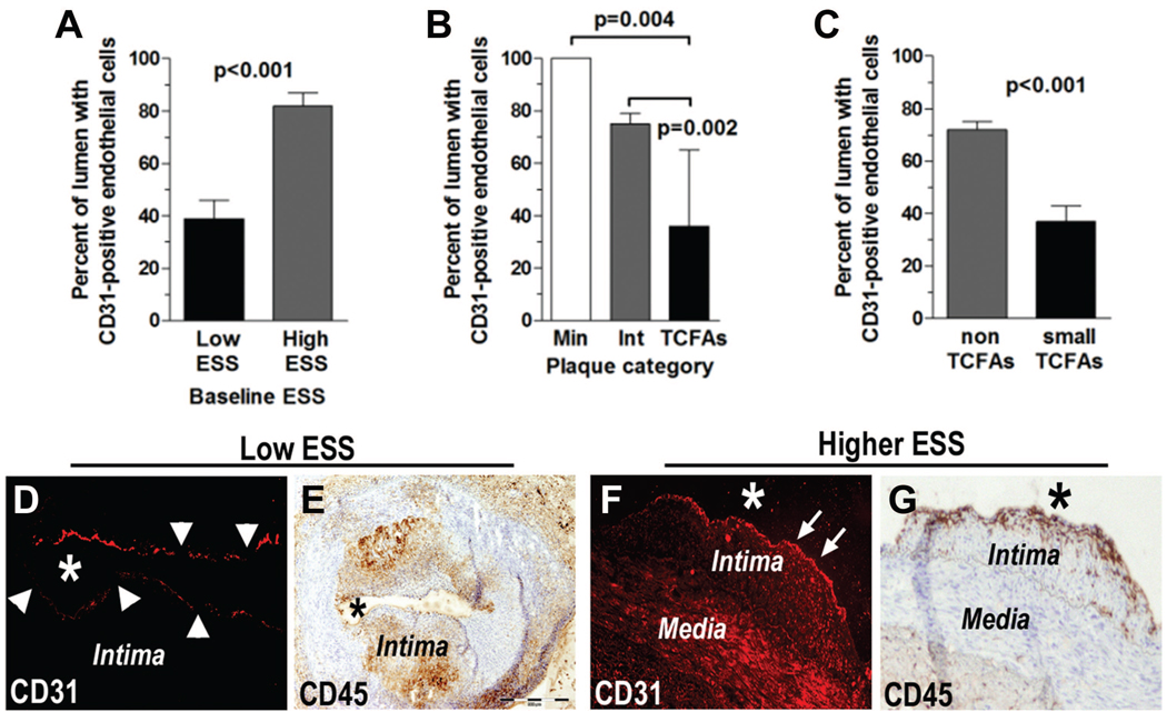
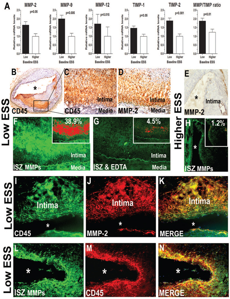
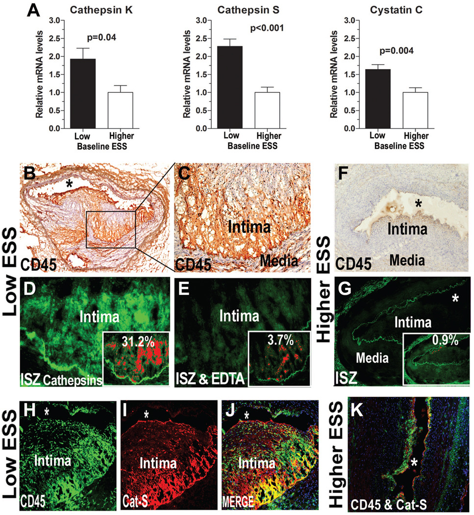
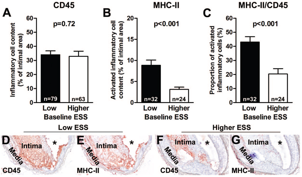
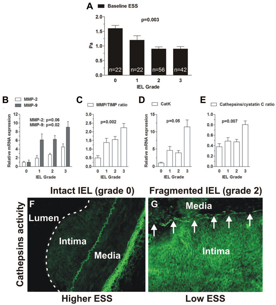
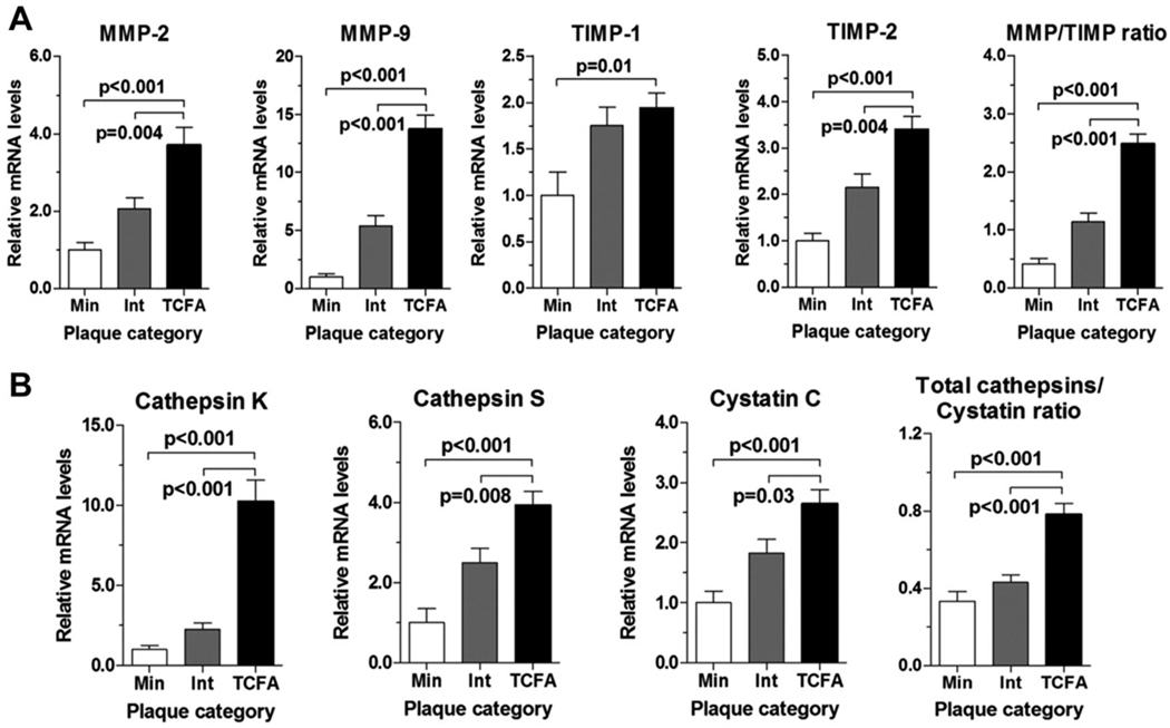
Background- The molecular mechanisms that determine the localized formation of thin-capped atheromata in the coronary arteries remain unknown. This study tested the hypothesis that low endothelial shear stress augments the expression of matrix-degrading proteases and thereby promotes the formation of thin-capped atheromata. Methods and Results- Intravascular ultrasound-based, geometrically correct 3-dimensional reconstruction of the coronary arteries of 12 swine was performed in vivo 23 weeks after initiation of diabetes mellitus and a hyperlipidemic diet. Local endothelial shear stress was calculated in plaque-free subsegments of interest (n=142) with computational fluid dynamics. At week 30, the coronary arteries (n=31) were harvested and the same subsegments were identified. The messenger RNA and protein expression and elastolytic activity of selected elastases and their endogenous inhibitors were assessed. Subsegments with low preceding endothelial shear stress at week 23 showed reduced endothelial coverage, enhanced lipid accumulation, and intense infiltration of activated inflammatory cells at week 30. These lesions showed increased expression of messenger RNAs encoding matrix metalloproteinase-2, -9, and -12, and cathepsins K and S relative to their endogenous inhibitors and increased elastolytic activity. Expression of these enzymes correlated positively with the severity of internal elastic lamina fragmentation. Thin-capped atheromata developed in regions with lower preceding endothelial shear stress and had reduced endothelial coverage, intense lipid and inflammatory cell accumulation, enhanced messenger RNA expression and elastolytic activity of MMPs and cathepsins, and severe internal elastic lamina fragmentation. Conclusions- Low endothelial shear stress induces endothelial discontinuity and accumulation of activated inflammatory cells, thereby augmenting the expression and activity of elastases in the intima and shifting the balance with their inhibitors toward matrix breakdown. Our results provide new insight into the mechanisms of regional formation of plaques with thin fibrous caps.


Figures

Figure 1
Figure 1
Percentage of luminal periphery with CD-31–positive endothelial cells in (A) low-ESS (n=18) vs higher-ESS (n=11) subsegments, (B) minimal lesions (Min) vs intermediate lesions (Int) vs atheromata with thin fibrous cap (TCFAs), and (C) small atheromata with thin fibrous cap (small TCFAs) vs similar-sized atheromata without fibrous caps (nonTCFAs). Representative (D) CD31 immunofluorescence and (E) CD45 immunostaining in a lesion with low baseline ESS show endothelial discontinuity (arrowheads) and intense inflammatory cell accumulation respectively. Representative (F) CD31 immunofluorescence and (G) CD45 immunostaining in a lesion of higher baseline ESS show intact endothelium (arrows) and limited accumulation of inflammatory cells respectively. Asterisks indicate the lumen.
Figure 2
Figure 2
Increased expression and activity of MMPs in low-ESS (n=79) vs higher-ESS (n=63) subsegments. A, Relative mRNA levels of MMP-2, MMP-9, MMP-12, TIMP-1, and TIMP-2 and the ratio of MMPs to TIMPs in low- vs higher-ESS subsegments. B, Representative immunostaining for CD45 in a low-ESS lesion. Higher magnification of (C) CD45 staining and (D) MMP-2 staining in the area selected by the black box in B. F, ISZ optimized for MMPs shows intense green fluorescence, indicating high elastolytic activity in serial sections with C and D. G, The MMP-inhibitor EDTA abolished elastolytic activity in the adjacent section. E, Absence of MMP-2 staining in a representative lesion of higher baseline ESS. H, Absence of MMP-mediated elastolytic activity in the same lesion as in E. Elastolytic activity–dependent fluorescence is shown in red in the insets in F through H. The percentage of intimal area with fluorescence intensity is indicated in each inset. I, Double immunofluorescence for (I) CD45 and (J) MMP-2 in a representative lesion of low ESS. K, Orange indicates colocalization of both antigens. L, Elastolytic activity (ISZ optimized for MMP) colocalizes with inflammatory cells infiltration (M, CD45 immunofluorescence applied in an adjacent section) in a lesion of low baseline ESS. N, Orange indicates colocalization. Asterisks indicate the lumen.
Figure 3
Figure 3
Increased expression and activity of cathepsins in low-ESS (n=79) vs higher-ESS (n=63) subsegments. A, Relative mRNA levels of cathepsins K and S and cystatin C in low- vs higher-ESS sub-segments. B, Representative immunostaining for CD45 in a lesion of low baseline ESS. C, Higher magnification of the region selected by the black box in B. D, ISZ optimized for cathepsins shows intense green fluorescence, indicating high elastolytic activity, in the inflammatory cell–rich area. E, Addition of cathepsin inhibitor E64D eliminates the enzyme activity in the serial section. F, CD45 immunostaining and (G) ISZ in a representative lesion of higher baseline ESS. Elastolytic activity–dependent fluorescence is shown in red in the insets in D, E, and G. The percentage of intimal area with fluorescence intensity is indicated in each inset. Immunofluorescent staining for (H) CD45 and (I) cathepsin S in a representative lesion of low ESS shows the marked expression and colocalization of both antigens, indicated by the merge of H and I (J). K, Double-immunofluorescent staining for CD45 (green) and cathepsin S (red) in a subsegment of higher ESS. Asterisks denote the lumen.
Figure 4
Figure 4
Low-ESS subsegments contain more activated inflammatory cells compared with higher ESS subsegments. A, The percentage of CD45-positive intimal area did not differ between low-ESS (n=79) and higher-ESS (n=63) subsegments. However, low-ESS sub-segments (n=32) had (B) a higher percentage of MCH-II–positive intima area and (C) a higher proportion of activated (MHC-II–positive) inflammatory cells compared with higher-ESS subsegments (n=24). Microphotographs represent (D) CD45 staining and (E) MCH-II staining in parallel sections of a low-ESS subsegment and a higher-ESS subsegment (F and G, respectively). Asterisks indicate the lumen.
Figure 5
Figure 5
The severity of IEL fragmentation correlates negatively with ESS levels and positively with MMP and cathepsin mRNA expression and activity. Association of each grade of IEL fragmentation with (A) baseline ESS, (B) relative mRNA expression of MMP-2 and MMP-9, (C) MMP/TIMP mRNA ratio, (D) relative mRNA expression of cathepsin K, and (E) cathepsins/cystatin C mRNA ratio. P values represent the overall association. In A through E, n=22 for IEL grade 1, n=22 for IEL grade 2, n=56 for IEL grade 3, and n=42 for IEL grade 4. Representative ISZ optimized for cathepsins in (F) a lesion of higher baseline ESS with intact IEL and (G) a lesion of low baseline ESS with moderate to severe IEL fragmentation; arrows depict the IEL breaks that colocalize with an increased elastolytic activity (bright green fluorescence) of cathepsins.
Figure 6
Figure 6
Relative mRNA levels of MMPs, cysteine proteases, and their inhibitors in minimal lesions (Min), intermediate lesions (Int), and atheromata with thin fibrous cap (TCFA). A, The mRNA levels of MMP-2, MMP-9, TIMP-1, and TIMP-2 and the total MMP/TIMP ratio. B, The mRNA levels of cathepsin K, cathepsin S, and cystatin C and the cathepsins/cystatin C ratio.

Similar articles

Cited by

References

    1. Chatzizisis YS, Coskun AU, Jonas M, Edelman ER, Feldman CL, Stone PH. Role of endothelial shear stress in the natural history of coronary atherosclerosis and vascular remodeling: molecular, cellular and vascular behavior. J Am Coll Cardiol. 2007;49:2379–2393. - PubMed
    1. Virmani R, Kolodgie FD, Burke AP, Farb A, Schwartz SM. Lessons from sudden coronary death: a comprehensive morphological classification scheme for atherosclerotic lesions. Arterioscler Thromb Vasc Biol. 2000;20:1262–1275. - PubMed
    1. Chatzizisis YS, Jonas M, Coskun AU, Beigel R, Stone BV, Maynard C, Gerrity R, Daley W, Campbell R, Elazer ER, Feldman CL, Stone PH. Prediction of the localization of high-risk coronary atherosclerotic plaques on the basis of low endothelial shear stress: an intravascular ultrasound and histopathology natural history study. Circulation. 2008;117:993–1002. - PubMed
    1. Koskinas KC, Feldman CL, Chatzizisis YS, Coskun AU, Jonas M, Maynard C, Baker AB, Edelman ER, Stone PH. Natural history of experimental coronary atherosclerosis and vascular remodeling in relation to endothelial shear stress: a serial, in vivo intravascular ultrasound study. Circulation. 2010;121:2092–2101. - PMC - PubMed
    1. Cheng C, Tempel D, van Haperen R, van der Baan A, Grosveld F, Daemen MJ, Krams R, de Crom R. Atherosclerotic lesion size and vulnerability are determined by patterns of fluid shear stress. Circulation. 2006;113:2744–2753. - PubMed

Publication types

MeSH terms