

Review
doi: 10.1161/HYPERTENSIONAHA.110.163519.
Epub 2011 Jan 31.
Intratubular renin-angiotensin system in hypertension
Affiliations
- PMID: 21282552
- PMCID: PMC3073668
- DOI: 10.1161/HYPERTENSIONAHA.110.163519
Item in Clipboard
Review
Intratubular renin-angiotensin system in hypertension
Hypertension.
2011 Mar.
No abstract available
Figures
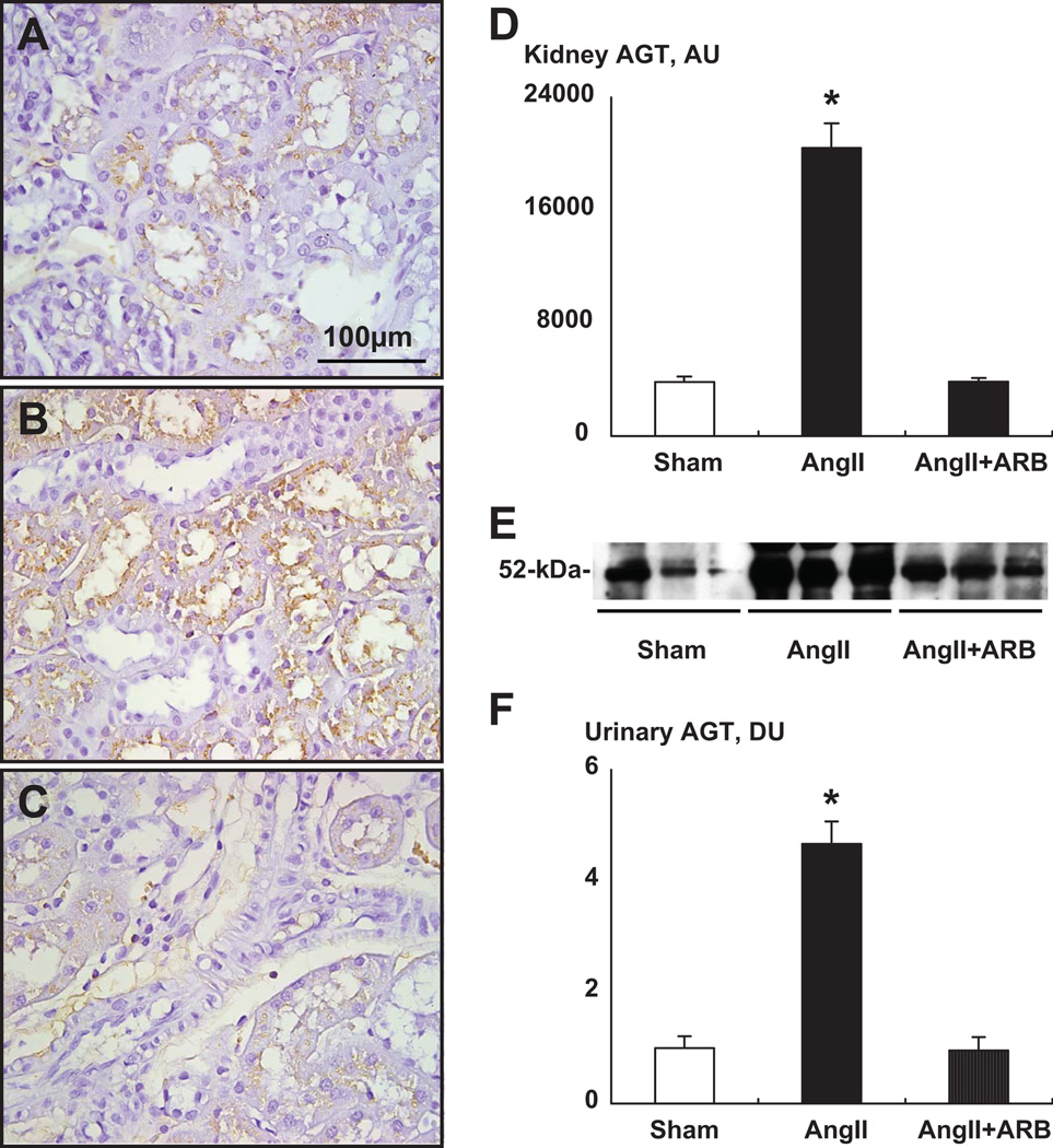
The immunoreactive areas were restricted only to proximal tubular cells. Vascular structures were negative. D, Kidney AGT immunostaining showed a significant enhancement in Ang II group (B) compared with sham group (A). ARB treatment prevented this augmentation (C). E, Representative Western blot analysis of urinary AGT levels among groups showing the stimulation in Ang II-infused group. F, Urinary excretion rates of AGT were enhanced 4.7-fold in Ang II-infused animals. ARB treatment prevented this augmentation. Ang II indicates angiotensin II; ARB, angiotensin II type1 receptor blocker, olmesartan; AGT, angiotensinogen. * P < 0.05 compared with the sham group. Data from Kobori et al. Hypertension 43:1126–1132, 2004.
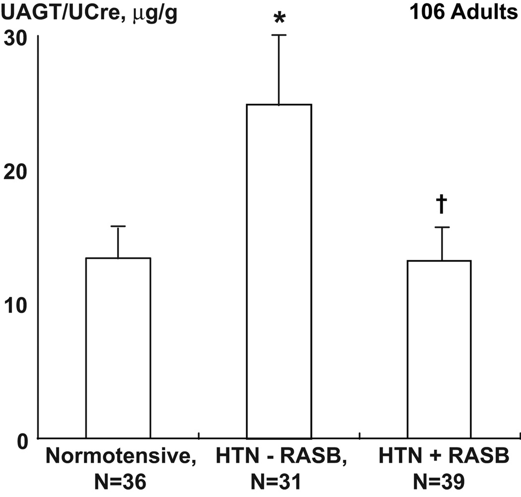
Uriinary AGT (uAGT), expressed as ratio of uAGT/uCreatine, in normotensive, and in hypertensive patients (HTN) treated with renin-angiotensin system blockers (RASB) and compared with those treated with other drugs. *P<0.05 vs normotensive; P<0.05 vs HTN–RASB. Data from Kobori et al. Hypertension 53[Part 2]:344–350, 2009.
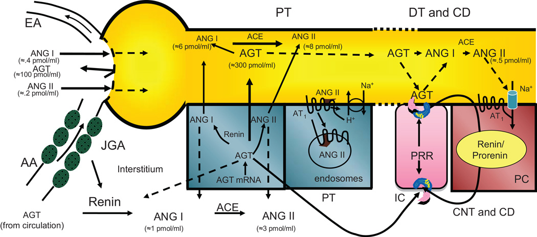
In Ang II dependent hypertension, the kidney maintains de novo intrarenal Ang II formation enhanced proximal tubule AGT formation and spillover into distal nephron segments coupled with enhancement of CD renin and stimulation of tubular ACE. (Refer to text for relevant references).
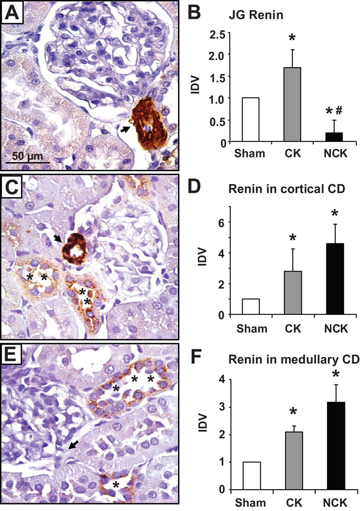
Renin immunoreactivity by immunoperoxidase technique (A,C, and E) in paraffin embedded kidney sections (3 µm) from sham rats (A), and clipped (C) and non-clipped kidney (E) of Goldblatt rats. Specific JG renin immunoreactivity (arrows; DAB chromogen) in a sham (A) and in the clipped kidney (C) of a Goldblatt rat. Higher renin immunoreactivity (asterisks) are shown in the collecting ducts of the renal cortexes of both, clipped (C) and non-clipped (E) kidneys relative to the sham kidney (A). Densitometry the renin immunoreactivity in JG cells (B) and cortical (D) and medullary (F) collecting duct cells of sham, and clipped (CK) and non-clipped (NCK) kidneys of Goldblatt rats were performed using four kidney sections/animal (10 microscopic fields/kidney sections at the renal cortex and medulla regions) and compared to sham kidneys. Sham rats (n= 5). Goldblatt rats (n= 6). Glom: Glomerulus. Values are mean ± S.E. *P<0.0001 versus sham. Renin antibody dilution 1:4,000. *P<0.05 versus sham. #P< 0.05 clipped kidney versus non-clipped. CD: collecting duct; JG: juxtaglomerular; CK: clipped kidney; NCK: non-clipped kidney; IDV: integrated densitometric values. Modified from Prieto-Carrasquero et al. Hypertension 51:1590–1596, 2008.
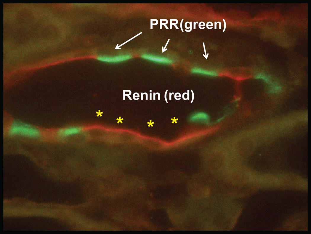
Double immunolabeling for renin (red) and (P)RR (green) was performed to confirm that renin is localized in principal cells (asterisks) while (P)RR is expressed in intercalated cells (arrows). A rabbit polyclonal anti-renin antibody (T. Inagami, Vanderbilt University) at a 1:4,000 dilution detected by a fluorescent secondary antibody (Alexa Fluor 594, red; Invitrogen) chicken anti-rabbit, was followed by a goat anti-(P)RR antibody (Abcam 5959, Cambridge, MA) at 1:400 dilution detected with a fluorescent secondary antibody donkey anti-goat (Alexa Fluor 488, green; Invitrogen). (Unpublished data)
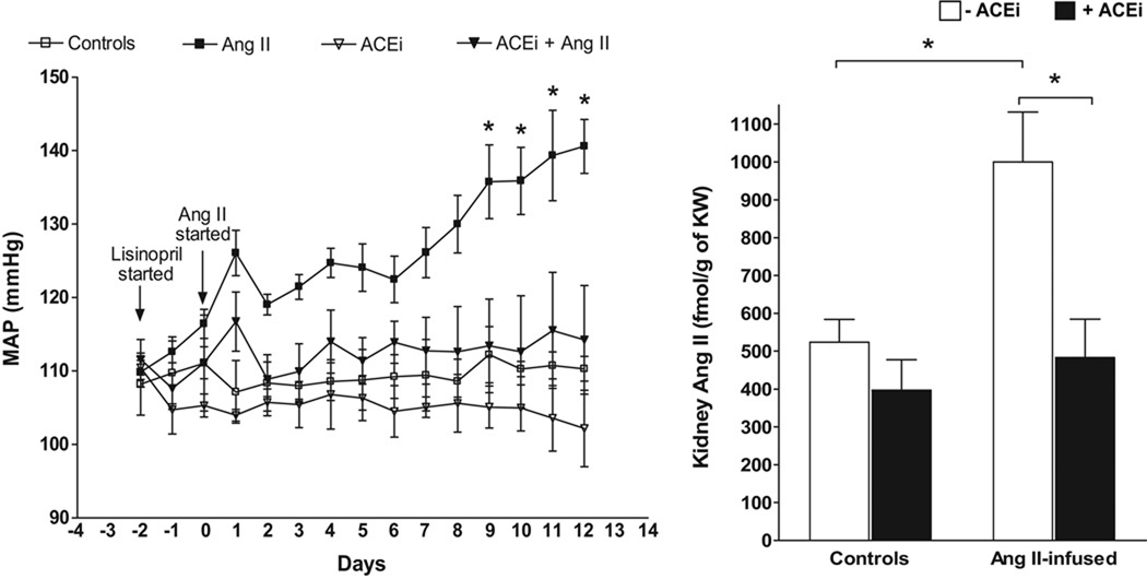
Blood pressure and Ang II concentrations were determined by telemetry and radioimmunoanalysis respectively. Ang II = Angiotensin II (400 ng/kg/min), ACEi = Lisinopril (100 mg/L in the drinking water). *p < 0.05 vs. controls by TWO-WAY ANOVA for MAP changes and ONE-WAY ANOVA for Ang II changes. (From Gonzalez-Villalobos et al. Hypertension 53:351–355, 2009).
References
-
- Kobori H, Nangaku M, Navar LG, Nishiyama A. The intrarenal renin-angiotensin system: from physiology to the pathobiology of hypertension and kidney disease. Pharmacol Rev. 2007;59:251–287. - PubMed
-
- Navar LG, Prieto-Carrasquero MC, Kobori H. Molecular aspects of the renal renin-angiotensin system. In: Re R, DiPette DJ, Schiffrin EL, Sowers JR, editors. Molecular Mechanisms in Hypertension. Taylor & Francis Group; 2006. pp. 3–14.
-
- Douglas JG, Hopfer U. Novel aspects of angiotensin receptors and signal transduction in the kidney. Annu Rev Physiol. 1994;56:649–669. - PubMed
-
- Navar LG, Harrison-Bernard LM, Imig JD, Mitchell KD. Renal actions of angiotensin II at AT1 receptor blockers. In: Epstein M, Brunner HR, editors. Angiotensin II Receptor Antagonists. Philadelphia: Hanley & Belfus, Inc.; 2000. pp. 189–214.
Publication types
MeSH terms
Substances
Grants and funding
LinkOut - more resources
Full Text Sources
Medical
