Review
. 2011 Mar 2;103(5):368-83.
doi: 10.1093/jnci/djq562. Epub 2011 Jan 31.

Human papillomavirus testing in the prevention of cervical cancer

Affiliations
Review

Human papillomavirus testing in the prevention of cervical cancer

Mark Schiffman et al. J Natl Cancer Inst. .

Abstract

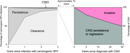
Strong evidence now supports the adoption of cervical cancer prevention strategies that explicitly focus on persistent infection with the causal agent, human papillomavirus (HPV). To inform an evidence-based transition to a new public health approach for cervical cancer screening, we summarize the natural history and cervical carcinogenicity of HPV and discuss the promise and uncertainties of currently available screening methods. New HPV infections acquired at any age are virtually always benign, but persistent infections with one of approximately 12 carcinogenic HPV types explain virtually all cases of cervical cancer. In the absence of an overtly persistent HPV infection, the risk of cervical cancer is extremely low. Thus, HPV test results predict the risk of cervical cancer and its precursors (cervical intraepithelial neoplasia grade 3) better and longer than cytological or colposcopic abnormalities, which are signs of HPV infection. The logical and inevitable move to HPV-based cervical cancer prevention strategies will require longer screening intervals that will disrupt current gynecologic and cytology laboratory practices built on frequent screening. A major challenge will be implementing programs that do not overtreat HPV-positive women who do not have obvious long-term persistence of HPV or treatable lesions at the time of initial evaluation. The greatest potential for reduction in cervical cancer rates from HPV screening is in low-resource regions that can implement infrequent rounds of low-cost HPV testing and treatment.


Figures

Figure 1
Figure 1
The cervical transformation zone and uncertainty of colposcopic impression. A) The tissue at risk of cervical cancer is the epithelial transformation zone, where the squamous vaginal epithelium undermines and replaces the glandular epithelium of the cervical canal. B) Squamous metaplasia continues as women age; effective screening, diagnosis, and treatment of the transformation zone become difficult. For women of any age, colposcopists cannot easily diagnose or target the biopsy of lesions of different underlying severity. Photographs were taken using a Zeiss 150 FC colposcope at 7.1-fold magnification. CIN1–3 = cervical intraepithelial neoplasia grades 1–3.
Figure 2
Figure 2
The evolution of human papillomavirus (HPV) types predicts carcinogenicity. The evolution of papillomaviruses is very slow. Tissue specificity, natural history, and carcinogenicity of HPVs are generally consistent with evolutionary relationships. HPV species in the alpha genus (left) infect mucosa, including the anogenital region and the oral cavity. The phylogenetic tree is based on the alignment of concatenated early and late open reading frames as described in Schiffman et al. (20). HPV types in the blue clade (which comprises species α1, α8, α10, and α13) include the HPV types that cause genital warts. HPV types in the green clade (which comprises species α2, α3, α4, and α15) cause commensal infections. HPV types in the red clade (which comprises species α5, α6, α7, α9, and α11) (shown in detail on the right) are associated to different degrees with cervical cancer and cervical intraepithelial neoplasia grade 3. The eight types that most commonly cause cervical cancer everywhere in the world belong to species alpha-9 or, to a lesser extent, to alpha-7. The carcinogen group for each HPV type is according to the International Agency for Research on Cancer (19): group 1 = carcinogenic, group 2A = probably carcinogenic, and group 2B = possibly carcinogenic.
Figure 3
Figure 3
Risks of human papillomavirus (HPV) persistence and progression. Left graph: Proportion of prevalent carcinogenic HPV infections that clear, persist, or progress to cervical intraepithelial neoplasia grade 3 (CIN3) in the first 3 years after first detection, based on all infections found at baseline screening in the Guanacaste Natural History Study (26). The great majority represented “new” infections (27). Persistence without CIN3 is surprisingly uncommon. Uncommon reappearances of HPV types following clearance did not predict risk of CIN3. Right graph: Proportion of untreated CIN3 lesions that invade to cancer within 30 years following the initial diagnosis [based on data from New Zealand (28)].
Figure 4
Figure 4
Cervical cancer progression model and optimized prevention strategy. A) The three steps in cervical carcinogenesis are acute human papillomavirus (HPV) infection, HPV persistence associated with the development of cervical precancer (cervical intraepithelial neoplasia grade 3 [CIN3] in particular, and invasion. B) Infection and clearance are extremely common and together contribute to the sharp peak of HPV prevalence in the years following average age of first sexual intercourse in the population. The average age at CIN3 diagnosis depends on the intensity of the screening effort in the population. Typically, it takes decades for a CIN3 lesion to grow and eventually invade, although there are rare and rapid exceptions. Panel B schematically displays current and future screening options integrating cytology (C), HPV testing (H), and prophylactic vaccination (V). The current emphasis on repeated cytology screening is inefficient. Even cotesting at 3-year intervals would not be optimal. At some point, an ideal program could emphasize HPV vaccination before average age at first sexual intercourse and reliance on increasingly spaced HPV tests for primary screening (or another equally sensitive method). Note: the prevalence curves are not drawn to scale. The graph shows age-related prevalence of HPV infections (green), CIN2 and CIN3 (blue), and cervical cancer (red) in the United States. Data on HPV infections are based on a summary of US-based HPV prevalence studies (55). The age distribution of CIN2 and/or CIN3 is based on data from Kaiser Permanente Northern California Health Maintenance Organization (P. E. Castle, personal communication) and the data on cancers are from the Surveillance, Epidemiology, and End Results 17 database (56).
Figure 5
Figure 5
Cumulative incidence rate of cervical intraepithelial neoplasia grade 3 or invasive cervical cancer (CIN3+) over 15 years following a single human papillomavirus (HPV) test. A cohort of 20 000 women from Kaiser Permanente (Portland, OR) was followed up by conventional cytology screening for approximately 15 years (78). Archived cervical specimens obtained from the women at enrollment (baseline) were tested for carcinogenic HPV types. The risk estimates, adjusted for loss to follow-up, show primarily that in this older cohort (average age approximately 35 years), a negative HPV test predicts very low risk of subsequent CIN3+. Baseline test positivity for HPV16, HPV18, or HPV31 was most strongly linked to subsequent CIN3+.
Figure 6
Figure 6
Negative impact of testing for HPV too frequently. We plot the results of HPV testing performed at two time points (at enrollment [t = 0] and at year 3) in women enrolled in the Guanacaste Natural History Study; disease ascertainment occurred annually. The cumulative incidence rate of cervical intraepithelial neoplasia grade 2 or worse (CIN2+) is shown for women testing positive and negative at enrollment and again for women testing positive and negative at year 3 among those who tested negative at enrollment (27). The risk stratification of the first HPV test is excellent, but the second test is less effective in detecting prevalent disease if performed too soon. Testing HPV positive for prevalent HPV infections predicts an elevated risk of CIN2+ (the treatment threshold) found before the screening at year 3, whereas testing HPV negative predicts a very low risk. Among women with an HPV-negative test at enrollment (t = 0), those who test HPV positive 3 years later (ie, at the currently recommended interval) are at much lower risk of CIN2+ than women who were HPV positive at the first screen and who may already have a persistent infection.
Figure 7
Figure 7
The use of risk bands to guide clinical decisions. Use of risk estimates simplifies a complex battery of test results over time into a single risk score on which management can be based. The probability of diagnosis of cervical intraepithelial neoplasia grade or cervical cancer (CIN3+) is a reasonable metric that can be calculated based on data from a prospective study. Importantly, various combinations of current test results, past test results, and human papillomavirus (HPV) vaccination history should result in similar management of women who, in fact, are at a similar risk of CIN3+. For example, the risk of CIN3+ for a woman who cotests HPV positive with atypical squamous cells of undetermined significance (ASC-US) equals that of a woman with low-grade squamous intraepithelial lesion (LSIL) by cytology; these women are referred to colposcopy. The risk of CIN3+ for a woman who tests HPV negative with ASC-US is similar to that for a woman who is HPV negative and has a normal cytology; these women should have follow-up screenings at similar intervals. New technologies, once validated in a clinical trial, would be integrated into the risk-based model, without the need for new consensus conferences on management. This graph is modified from Castle et al. (95). HSIL = high-grade squamous intraepithelial lesion.

Comment in

References

    1. Gustafsson L, Pontén J, Bergström R, Adami HO. International incidence rates of invasive cervical cancer before cytological screening. Int J Cancer. 1997;71(2):159–165. - PubMed
    1. Gustafsson L, Pontén J, Zack M, Adami HO. International incidence rates of invasive cervical cancer after introduction of cytological screening. Cancer Causes Control. 1997;8(5):755–763. - PubMed
    1. Saslow D, Runowicz CD, Solomon D, et al. American Cancer Society guideline for the early detection of cervical neoplasia and cancer. CA Cancer J Clin. 2002;52(6):342–362. - PubMed
    1. Schiffman M, Castle PE, Jeronimo J, Rodriguez AC, Wacholder S. Human papillomavirus and cervical cancer. Lancet. 2007;370(9590):890–907. - PubMed
    1. International Agency for Research on Cancer. Cervix Cancer Screening. Lyon, France: IARC Press; 2005.

Publication types

MeSH terms