. 2011 Feb 15;108(7):2795-800.
doi: 10.1073/pnas.1013124108. Epub 2011 Jan 31.

Nε-lysine acetylation determines dissociation from GAP junctions and lateralization of connexin 43 in normal and dystrophic heart

Affiliations

Nε-lysine acetylation determines dissociation from GAP junctions and lateralization of connexin 43 in normal and dystrophic heart

Claudia Colussi et al. Proc Natl Acad Sci U S A. .

Abstract

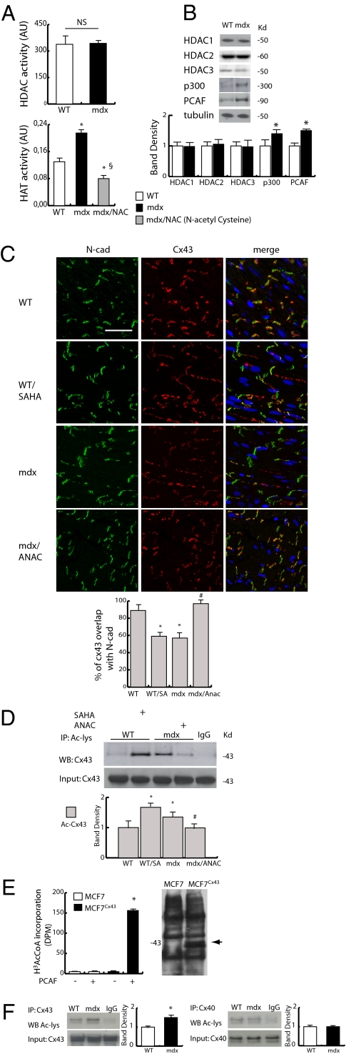
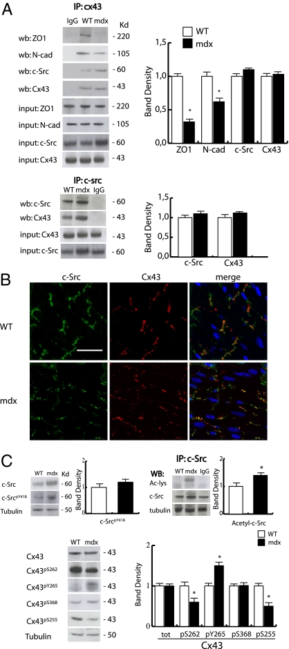
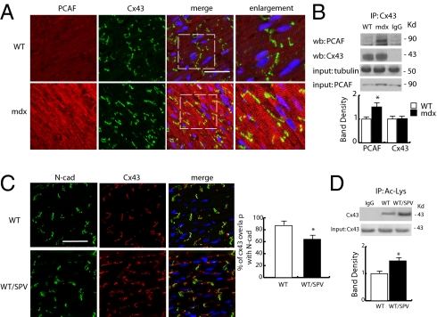
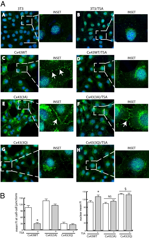
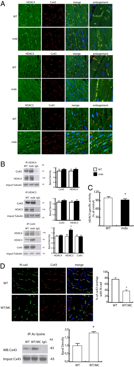
Wanting to explore the epigenetic basis of Duchenne cardiomyopathy, we found that global histone acetylase activity was abnormally elevated and the acetylase P300/CBP-associated factor (PCAF) coimmunoprecipitated with connexin 43 (Cx43), which was N(ε)-lysine acetylated and lateralized in mdx heart. This observation was paralleled by Cx43 dissociation from N-cadherin and zonula occludens 1, whereas pp60-c-Src association was unaltered. In vivo treatment of mdx with the pan-histone acetylase inhibitor anacardic acid significantly reduced Cx43 N(ε)-lysine acetylation and restored its association to GAP junctions (GJs) at intercalated discs. Noteworthy, in normal as well as mdx mice, the class IIa histone deacetylases 4 and 5 constitutively colocalized with Cx43 either at GJs or in the lateralized compartments. The class I histone deacetylase 3 was also part of the complex. Treatment of normal controls with the histone deacetylase pan-inhibitor suberoylanilide hydroxamic acid (MC1568) or the class IIa-selective inhibitor 3-{4-[3-(3-fluorophenyl)-3-oxo-1-propen-1-yl]-1-methyl-1H-pyrrol-2-yl}-N-hydroxy-2-propenamide (MC1568) determined Cx43 hyperacetylation, dissociation from GJs, and distribution along the long axis of ventricular cardiomyocytes. Consistently, the histone acetylase activator pentadecylidenemalonate 1b (SPV106) hyperacetylated cardiac proteins, including Cx43, which assumed a lateralized position that partly reproduced the dystrophic phenotype. In the presence of suberoylanilide hydroxamic acid, cell to cell permeability was significantly diminished, which is in agreement with a Cx43 close conformation in the consequence of hyperacetylation. Additional experiments, performed with Cx43 acetylation mutants, revealed, for the acetylated form of the molecule, a significant reduction in plasma membrane localization and a tendency to nuclear accumulation. These results suggest that Cx43 N(ε)-lysine acetylation may have physiopathological consequences for cell to cell coupling and cardiac function.


Conflict of interest statement

The authors declare no conflict of interest.

Figures

Fig. 1.
Fig. 1.
Evaluation of epigenetic enzymes expression and Cx43 acetylation. (A Upper) Evaluation of HDACs activity in WT and mdx heart. (Lower) HAT activity in normal (WT), mdx, and mdx mice treated with N-acetyl cysteine (NAC). *P < 0.05 vs. WT; §P < 0.05 vs. mdx. (B) Western blotting analysis of HDAC1, -2, and -3, p300, and PCAF expression in WT and mdx heart. Densitometry is shown in Lower (P < 0.05; n = 3). (C Upper) Confocal analysis of Cx43 (red) and N-cad (green) distribution in a heart section from WT, WT treated with SAHA, mdx, and mdx treated with anacardic acid (ANAC) mice. Nuclei were counterstained with 157199-63-8/Quinolinium, 4-{3-[3-methyl-2(3H)-benzothiazolylidene]-1-propenyl}-1-[3-(trimethylammonio)propyl]-,diiodide (TOPRO3) (blue). (Scale bar, 50 μm.) (Lower) The graph shows the degree of Cx43 and N-cad colocalization (%) at intercalated discs (ICDs). (D) Western blotting analysis of Cx43 acetylation level in WT, WT treated with SAHA, mdx, and mdx treated with ANAC mice. Densitometric analysis is shown in Lower. *P < 0.05 vs. WT; #P < 0.05 vs. mdx. (E) H3-acetyl-CoA in mock or cytomegalovirus promoter (pCMV)-Cx43–transfected MFC7 cells (MCF7Cx43). Assays were performed in the presence or absence of recombinant PCAF added to the fresh lysate. Right shows Cx43 expression in mock and pCMV-Cx43 transfected MFC7 cells. (F) Immunoprecipitation and Western blotting analysis of Cx43 and connexin 40 (Cx40) expression and acetylation in the WT and mdx heart.
Fig. 2.
Fig. 2.
Characterization of Cx43 phosphorylation and binding partners association. (A Upper) Immunoprecipitation analysis showing the association level of N-cad, ZO1, and c-Src with Cx43 in WT and mdx heart. (Lower) Association between c-Src and Cx43 in WT and mdx heart. Densitometric analyses are shown in Right. *P < 0.05. (B) Confocal analysis of c-Src (green) and Cx43 (red) distribution in WT and mdx heart. Nuclei were counterstained with TOPRO3 (blue). (Scale bar, 50 μm.) (C) Western blotting analysis showing phosphorylation level of c-Src (Y418) and Cx43 (S262, Y265, S368, and S255). Right shows band density. *P < 0.05.
Fig. 3.
Fig. 3.
Evaluation of PCAF distribution and function. (A) Panels show representative confocal images depicting PCAF (red) and Cx43 (green) expression and distribution in WT and mdx heart. Nuclei were counterstained with TOPRO3 (blue). (Scale bar, 50 μm.) The far right column shows enlarged details identified by brackets. (Scale bar, 50 μm.) (B Upper) Coimmunoprecipitation analysis of PCAF and Cx43 in the mdx heart. (Lower) Densitometric analysis. *P < 0.05. (C) Confocal analysis of N-cad (green) and Cx43 (red) distribution from control and SPV106-treated WT mice hearts. Nuclei were counterstained with TOPRO3 (blue). (Scale bar, 50 μm.) Right shows percentage of Cx43 and N-cad overlapping at ICDs level. (D) Immunoprecipitation experiment showing increase in Cx43 acetylation in WT treated with SPV106 or equivalent amount of solvent. Densitometric analysis is shown on the right. *P < 0.05.
Fig. 4.
Fig. 4.
Acetylation alters Cx43 intracellular distribution. (A) Confocal analysis of NIH 3T3 cells transient-transfected with mock (A and B), WT (Cx43WT; C and D), or K → A (Cx433A; E and F) and K → Q (Cx433Q; G and H) connexin 43 expression vectors. Samples were analyzed before (A, C, D, and G) and after TSA treatment (B, D, F, and H). Insets show enlarged details representative of each experimental condition. Transfection efficiency was 80% on average for each vector. Experiments were repeated three times. (B) Quantitative evaluation of Cx43WT, Cx433A, and Cx433Q distribution at cell to cell junctions (Left) or the nuclear level (Right). Data are expressed as mean fluorescence intensity (MFI); about 300 cells were counted for each experimental condition. *P < 0.05 vs. Cx43WT in untreated cells; §P < 0.05 vs. Cx43WT in untreated cells.
Fig. 5.
Fig. 5.
HDACs localization, function, and association with Cx43. (A) Confocal analysis of Cx43 (red) and HDAC4 (green; Top), HDAC5 (green; Middle), or HDAC3 (green; Bottom) expression and distribution in the WT and mdx heart. Nuclei were counterstained with TOPRO3 (blue). (Scale bar, 50 μm.) Enlarged details are shown in the far right column. (Scale bar, 50 μm.) (B) Coimmunoprecipitation experiments showing HDAC4, -5, and -3 association with Cx43 in the WT and mdx heart. Relative densitometries are shown in Right. *P < 0.05. (C) HDAC4-specific activity evaluated in the WT and mdx heart samples. *P < 0.05. (D) Confocal analysis of N-cad (green) and Cx43 (red) expression and distribution in hearts from WT treated with MC1568 or equivalent amounts of solvent. Nuclei were counterstained with TOPRO3 (blue). (Scale bar, 50 μm.) The graph shows Cx43 and N-cad percentage of colocalization. *P < 0.05. Lower shows the acetylation level of Cx43 in WT treated with MC1568 or an equivalent amount of solvent (Left). Densitometry is shown to Right. *P < 0.05.

References

    1. Beggs AH. Dystrophinopathy, the expanding phenotype. Dystrophin abnormalities in X-linked dilated cardiomyopathy. Circulation. 1997;95:2344–2347. - PubMed
    1. Bia BL, et al. Decreased myocardial nNOS, increased iNOS and abnormal ECGs in mouse models of Duchenne muscular dystrophy. J Mol Cell Cardiol. 1999;31:1857–1862. - PubMed
    1. Wehling-Henricks M, Jordan MC, Roos KP, Deng B, Tidball JG. Cardiomyopathy in dystrophin-deficient hearts is prevented by expression of a neuronal nitric oxide synthase transgene in the myocardium. Hum Mol Genet. 2005;14:1921–1933. - PubMed
    1. Williams IA, Allen DG. The role of reactive oxygen species in the hearts of dystrophin-deficient mdx mice. Am J Physiol Heart Circ Physiol. 2007;293:H1969–H1977. - PubMed
    1. Hitchler MJ, Domann FE. An epigenetic perspective on the free radical theory of development. Free Radic Biol Med. 2007;43:1023–1036. - PMC - PubMed

Publication types