. 2011 Feb 15;108(7):2909-14.
doi: 10.1073/pnas.1018892108. Epub 2011 Jan 31.

Losartan inhibits collagen I synthesis and improves the distribution and efficacy of nanotherapeutics in tumors

Affiliations

Losartan inhibits collagen I synthesis and improves the distribution and efficacy of nanotherapeutics in tumors

Benjamin Diop-Frimpong et al. Proc Natl Acad Sci U S A. .

Abstract

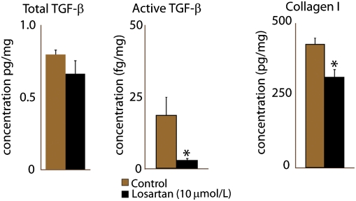
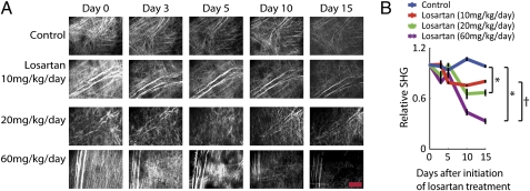
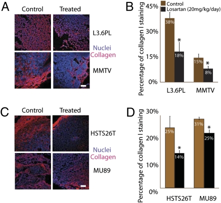
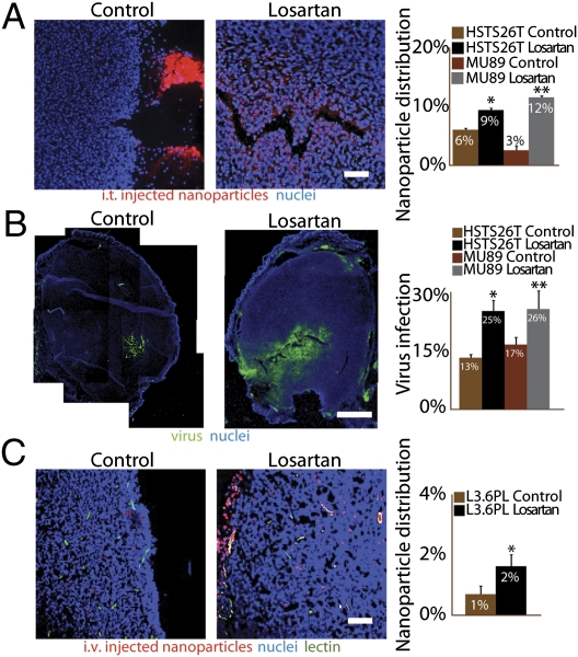
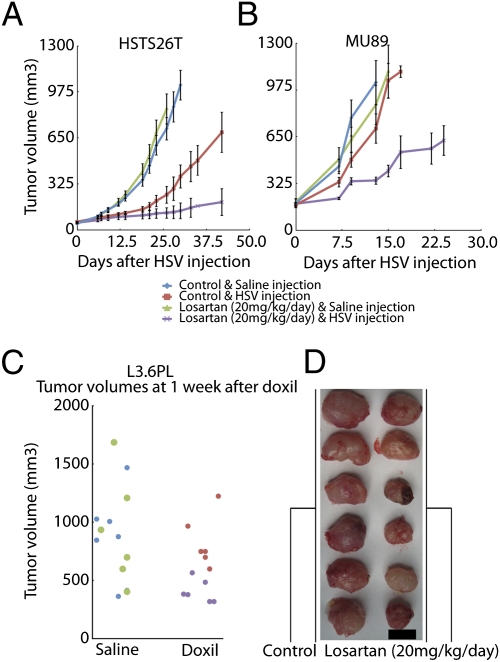
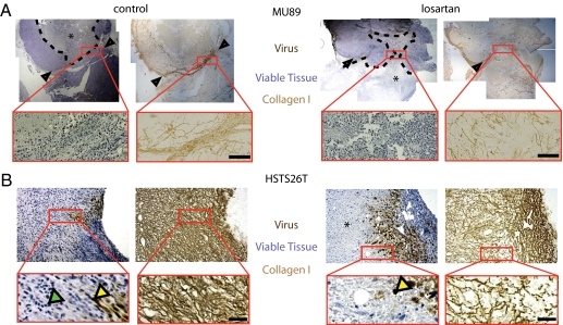
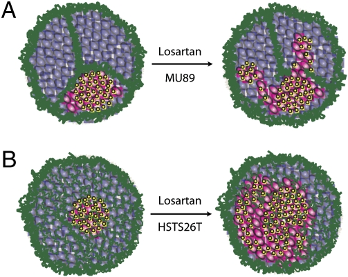
The dense collagen network in tumors significantly reduces the penetration and efficacy of nanotherapeutics. We tested whether losartan--a clinically approved angiotensin II receptor antagonist with noted antifibrotic activity--can enhance the penetration and efficacy of nanomedicine. We found that losartan inhibited collagen I production by carcinoma-associated fibroblasts isolated from breast cancer biopsies. Additionally, it led to a dose-dependent reduction in stromal collagen in desmoplastic models of human breast, pancreatic, and skin tumors in mice. Furthermore, losartan improved the distribution and therapeutic efficacy of intratumorally injected oncolytic herpes simplex viruses. Finally, it also enhanced the efficacy of i.v. injected pegylated liposomal doxorubicin (Doxil). Thus, losartan has the potential to enhance the efficacy of nanotherapeutics in patients with desmoplastic tumors.


Conflict of interest statement

Conflict of interest statement: R.K.J. received commercial research grants from Dyax, AstraZeneca, and MedImmune; consultant fees from AstraZeneca/MedImmune, Dyax, Astellas-Fibrogen, Regeneron, Genzyme, Morphosys, and Noxxon Pharma; and a speaker honorarium from Genzyme. R.K.J. owns stock in SynDevRx. No reagents or funding from these companies was used in these studies. There is no significant financial or other competing interest in the work.

Figures

Fig. 1.
Fig. 1.
Losartan reduces TGF-β1 activation and collagen I production in carcinoma-associated fibroblasts in vitro. Cells were treated with 10 μmol/L losartan for 24 h. Losartan reduced by 90% the active TGF-β1 levels, whereas total TGF-β1 levels were unaffected. There was a corresponding 27% decrease in collagen I levels. The reduction in active TGF-β1 and collagen I was statistically significant (Student's t test, *P < 0.05).
Fig. 2.
Fig. 2.
Losartan reduces collagen production in tumors. (A) Over a period of 2 wk, there was a dose-dependent reduction in collagen levels assessed by SHG imaging in losartan-treated HSTS26T tumors (10, 20, and 60 mg·kg−1·d−1). (Scale bar, 200 μm.) (B) At the end of 15 d, losartan doses of 10, 20, and 60 mg·kg−1·d−1 decreased the SHG levels by 20%, 33%, and 67%, respectively. There was a statistically significant difference (*) between the control group and the two higher doses (20 and 60 mg·kg−1·d−1). There was also a statistically significant difference (†) between the 20 and 60 mg·kg−1·d−1 groups.
Fig. 3.
Fig. 3.
Losartan reduces collagen levels in tumors. (A) Collagen I (red) and nuclei (blue) immunostaining in tumor sections in L3.6pl and MMTV control and losartan (20 mg·kg−1·d−1)-treated tumors. (Scale bar, 100 μm.) (B) The 2-wk losartan treatment at 20 mg·kg−1·d−1 significantly reduced the collagen I immunostaining in L3.6pl and FVB MMTV PyVT by 50% (*P < 0.03) and 47% (*P < 0.05), respectively. (C) Collagen I (red) and nuclei (blue) immunostaining in tumor sections in HSTS26T and Mu89 control and losartan (20 mg·kg−1·d−1)-treated tumors. Note that there is no reduction in collagen I immunostaining at 200 μm from the edge of HSTS26T tumors. This phenomenon is less obvious in treated Mu89 tumors, where there is some persistent staining both at the edge and in central tumor areas. (Scale bar, 100 μm.) (D) Losartan significantly reduced the collagen I immunostaining in HSTS26T and Mu89 by 42% (*P < 0.02) and 20% (*P < 0.05), respectively.
Fig. 4.
Fig. 4.
Losartan increases delivery of nanoparticles and nanotherapeutics. (A) Distribution of i.t. injected 100-nm-diameter nanoparticles in HSTS26T tumors. Losartan significantly increased (*, **P < 0.001) the distribution of i.t. injected nanoparticles in both tumor types (1.5-fold in HSTS26T and 4-fold in Mu89). An analysis of the distribution pattern shows control tumors with fewer intratumoral nanoparticles (red) and a majority of nanoparticles that backtracked out of the needle track and accumulated at the tumor surface. In contrast, treated tumors have a significant number of intratumoral nanoparticles. (Scale bar, 100 μm.) (B) Distribution of viral infection 24 h after the intratumoral injection of HSV-expressing green fluorescent protein. HSV infection (green) in control tumors is limited to the cells in close proximity to the injection site, whereas losartan-treated tumors have a more extensive spread of HSV infection. (Scale bar, 1 mm.) Losartan significantly increased (*, **P < 0.05) the virus spread in HSTS26T and Mu89 tumors. (C) Distribution of i.v. injected 100-nm-diameter nanoparticles in L3.6pl tumors. The nanoparticles (red) are localized around perfused vessels (green). There is a twofold increase (*P < 0.05) in nanoparticle content in losartan-treated tumors compared with control tumors. (Scale bar, 100 μm.)
Fig. 5.
Fig. 5.
Losartan significantly delays the growth of tumors treated with Doxil or HSV. (A and B) Mice bearing HSTS26T (A) and Mu89 (B) tumors were treated for 2 wk with either losartan or saline before the i.t. injection of HSV. Losartan alone did not affect the growth of Mu89 or HSTS26T tumors. The growth delay was significantly longer (P < 0.001) in HSTS26T tumors treated with losartan and HSV compared with tumors treated with HSV alone. The i.t. injection of HSV did not delay the growth of Mu89 tumors, but the combined losartan and HSV treatment significantly retarded (P < 0.001) the growth of Mu89 tumors. (C) Mice that received losartan treatment before i.v. Doxil infusion (purple dots) have significantly smaller (P < 0.001) tumors than those that received Doxil alone (red dots) in L3.6pl tumors. Note that there is no difference in tumor size between saline- (blue) and losartan-treated (green) mice. (D) The image shows a clear difference in size between control tumors (left column) and losartan-treated tumors (right column) at 1 wk after Doxil infusion. (Scale bar, 1 cm.)
Fig. 6.
Fig. 6.
Relationship between collagen structure and virus infection and necrosis. (A) In Mu89 tumors, collagen bundles are seen around the tumor margin. Occasionally, these bundles project into the tumor (black arrowheads) and divide the tumor into separate compartments. These compartments seem to confine movement of HSV, evident from the containment of the necrotic region (*) within the region bounded by collagen bundles. When these tumors were treated with losartan, the collagen bundles at the margins of the tumor remained intact but the projections became less organized (inset). This presumably allowed virus propagation and necrosis to extend across the boundaries. (Scale bar, 100 μm.) (B) In HSTS26T tumors, the dense mesh-like collagen network confined virus infection to the immediate area surrounding the injection point. With losartan treatment, there was a reduction in the density of the network that presumably allowed virus particles to infect a larger area and thus more tumor cells. Green and yellow arrowheads indicate viable and virus-infected cells, respectively. (Scale bar, 40 μm.)
Fig. 7.
Fig. 7.
Schematic of virus distribution and infection in Mu89 and HSTS26T tumors. The schematics show how the different collagen network structures affect virus propagation and distribution. The collagen fibers (green) restrict the movement of virus particles (yellow) and the infection (pink) of noninfected (purple) cancer cells. (A) In Mu89 tumors, collagen bundles divide the tumor into isolated regions that cannot be traversed by virus particles. Losartan treatment destabilizes the collagen bundles and allows virus particles to move from one region to another. (B) In HSTS26T tumors, the collagen structure is a mesh-like sieve. Virus particles can still propagate through the sieve but do not extend very far from the injection site. Losartan treatment significantly destabilizes the mesh structure in the internal regions of the tumor and allows the virus to propagate and infect a larger area.

References

    1. Jain RK, Stylianopoulos T. Delivering nanomedicine in solid tumors. Nat Rev Clin Oncol. 2010;7:653–664. - PMC - PubMed
    1. Davis ME, Chen ZG, Shin DM. Nanoparticle therapeutics: An emerging treatment modality for cancer. Nat Rev Drug Discov. 2008;7:771–782. - PubMed
    1. Peer D, et al. Nanocarriers as an emerging platform for cancer therapy. Nat Nanotechnol. 2007;2:751–760. - PubMed
    1. Torchilin VP. Recent advances with liposomes as pharmaceutical carriers. Nat Rev Drug Discov. 2005;4:145–160. - PubMed
    1. Netti PA, Berk DA, Swartz MA, Grodzinsky AJ, Jain RK. Role of extracellular matrix assembly in interstitial transport in solid tumors. Cancer Res. 2000;60:2497–2503. - PubMed

Publication types

MeSH terms