. 2011 Feb 15;108(7):2741-6.
doi: 10.1073/pnas.1016498108. Epub 2011 Jan 31.

XMAP215 polymerase activity is built by combining multiple tubulin-binding TOG domains and a basic lattice-binding region

Affiliations

XMAP215 polymerase activity is built by combining multiple tubulin-binding TOG domains and a basic lattice-binding region

Per O Widlund et al. Proc Natl Acad Sci U S A. .

Abstract

XMAP215/Dis1 family proteins positively regulate microtubule growth. Repeats at their N termini, called TOG domains, are important for this function. While TOG domains directly bind tubulin dimers, it is unclear how this interaction translates to polymerase activity. Understanding the functional roles of TOG domains is further complicated by the fact that the number of these domains present in the proteins of different species varies. Here, we take advantage of a recent crystal structure of the third TOG domain from Caenorhabditis elegans, Zyg9, and mutate key residues in each TOG domain of XMAP215 that are predicted to be important for interaction with the tubulin heterodimer. We determined the contributions of the individual TOG domains to microtubule growth. We show that the TOG domains are absolutely required to bind free tubulin and that the domains differentially contribute to XMAP215's overall affinity for free tubulin. The mutants' overall affinity for free tubulin correlates well with polymerase activity. Furthermore, we demonstrate that an additional basic region is important for targeting to the microtubule lattice and is critical for XMAP215 to function at physiological concentrations. Using this information, we have engineered a "bonsai" protein, with two TOG domains and a basic region, that has almost full polymerase activity.


Conflict of interest statement

The authors declare no conflict of interest.

Figures

Fig. 1.
Fig. 1.
XMAP215 and its homologues. (A) The three major classes of TOG proteins. The human, frog, fly, plant, worm, and yeast homologues are shown. (B) Positions of the point mutations in XMAP215TOG1-5AA.
Fig. 2.
Fig. 2.
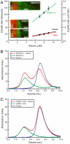
XMAP215TOG1-5AA-GFP does not promote microtubule growth and does not bind tubulin. (A) XMAP215-GFP shows a fivefold increase in tubulin growth at all tubulin concentrations examined while XMAP215TOG1-5AA-GFP does not. Error bars represent SEM (N≥15 for each point). Kymographs show a representative single microtubule from each experiment. Trend lines project back to the critical concentration in the absence of catastrophes. Rhodamine-labeled GMPCPP seeds are in red. Alexa488 labeled tubulin is in green. (B) XMAP215-GFP binds tubulin. Chromatography experiments showing A280 over elution volume. Three traces are shown in each graph: XMAP215-GFP alone in green, tubulin alone in blue, XMAP215-GFP in combination with tubulin in red. (C) XMAP215TOG1-5AA-GFP does not bind tubulin. Chromatography experiments showing A280 over elution volume. Three traces are shown in each graph: XMAP215TOG1-5AA-GFP alone in green, tubulin alone in blue, XMAP215-GFP in combination with tubulin in red.
Fig. 3.
Fig. 3.
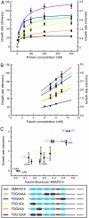
Characterization of all XMAP215 TOG domain point mutants. (A) Microtubule growth rate by XMAP215-GFP and all point mutants with increasing XMAP215 concentration at 5 μM tubulin. Error bars represent SEM (N≥10 for each point). (B) Growth promotion by XMAP215-GFP and all point mutants with increasing tubulin concentration. Error bars represent SEM (N≥10 for each point). (C) Maximum average growth rate of individual XMAP215 mutants plotted against their ability to bind tubulin as determined from chromatograms (Figs. S1 and S2). The maximum average growth rate was determined in at least four separate experiments at either 200 or 400 nM protein. Because no significant difference was seen between 200 and 400 nM protein for each construct, the rates were averaged (N≥10 for each experiment). Error bars represent SEM. Tubulin binding was measured in duplicate.
Fig. 4.
Fig. 4.
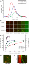
Construction of a minimal polymerase. (A) TOG12 binds tubulin. Chromatography experiments showing A280 over elution volume. Three traces are shown in each graph: TOG12 alone in green, tubulin alone in blue, TOG12 in combination with tubulin in red. (B) The Kif2A lattice-binding domain increases affinity of TOG12 for the microtubule lattice. Fragments were incubated with rhodamine-labeled GMPCPP-stabilized microtubules with increasing protein concentration as indicated. The merged image shows microtubules in red and GFP in green. (C) Microtubule growth rate with increasing TOG12, TOG12+, and TOG12+++ concentration at 5 μM tubulin. Error bars represent SD (N≥10 for each point). (D) TOG12 binds the plus end of stabilized GMPCPP seeds. Rhodamine-labeled GMPCPP seeds are in red. TOG12-GFP is in green. (E) TOG12+++ GFP tracks growing microtubule plus ends. Rhodamine-labeled GMPCPP seeds are in red. TOG12+++GFP is in green.
Fig. 5.
Fig. 5.
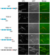
The microtubule-lattice-binding domain lies between TOG4 and TOG5. 200 nM XMAP215-GFP and various deletion mutants were incubated with rhodamine-labeled GMPCPP-stabilized microtubules and imaged. The left image shows the microtubules. The center image shows the GFP signal. The right image shows the merged image with microtubules in red and GFP in green.
Fig. 6.
Fig. 6.
XMAP215 as a catalyst. (A) Mutation or removal of TOG domains result in mutants that have lowered maximal growth rates (νmax) at any fixed tubulin concentration. The graph shows theoretical dose response of a protein with increasing affinities for the tubulin dimer that lead to increasing affinities for the transition state (compared to the microtubule end alone): α = 2 in red, α = 5 in green, and α = 10 in blue. β = 1, [T] = 0.1K1 (see SI Text). (B) Mutation or removal of the microtubule-binding domain in XMAP215 results in constructs that have the same νmax but a higher KD. The graph shows theoretical dose response of a protein with a constant νmax and an altered kon for microtubule-lattice binding: 4x reduced in red, 2x reduced in green, not reduced in blue. β = 1, [T] = 0.1K1 (see SI Text). (C) Model of TOG12+++ on the plus end of a microtubule. (1) Diffusion to the end via the lattice-binding domain. (2) TOG12+++ stabilizes the incoming dimer. (3) TOG12+++ remains bound to the incorporated dimer. (4) Release of the dimer. The transitions between each state are described by the reaction scheme in the SI Text.

References

    1. Howard J, Hyman AA. Microtubule polymerases and depolymerases. Curr Opin Cell Biol. 2007;19:31–35. - PubMed
    1. Pantaloni D, Le Clainche C, Carlier MF. Mechanism of actin-based motility. Science. 2001;292:1502–1506. - PubMed
    1. Desai A, Mitchison TJ. Microtubule polymerization dynamics. Annu Rev Cell Dev Biol. 1997;13:83–117. - PubMed
    1. Paul A, Pollard T. Review of the mechanism of processive actin filament elongation by formins. Cell Motil Cytoskeleton. 2009;66:606–617. - PMC - PubMed
    1. Goode BL, Drubin DG, Barnes G. Functional cooperation between the microtubule and actin cytoskeletons. Curr Opin Cell Biol. 2000;12:63–71. - PubMed

Publication types

MeSH terms

LinkOut - more resources