. 2011 Feb 15;108(7):2813-8.
doi: 10.1073/pnas.1012090108. Epub 2011 Jan 31.

Dishevelled is essential for neural connectivity and planar cell polarity in planarians

Affiliations

Dishevelled is essential for neural connectivity and planar cell polarity in planarians

Maria Almuedo-Castillo et al. Proc Natl Acad Sci U S A. .

Abstract

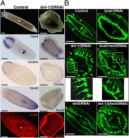
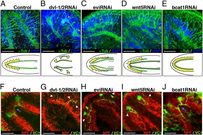
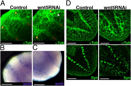
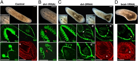
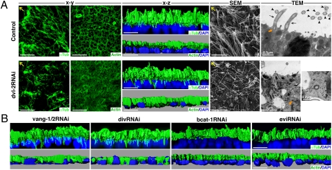
The Wingless/Integrated (Wnt) signaling pathway controls multiple events during development and homeostasis. It comprises multiple branches, mainly classified according to their dependence on β-catenin activation. The Wnt/β-catenin branch is essential for the establishment of the embryonic anteroposterior (AP) body axis throughout the phylogenetic tree. It is also required for AP axis establishment during planarian regeneration. Wnt/β-catenin-independent signaling encompasses several different pathways, of which the most extensively studied is the planar cell polarity (PCP) pathway, which is responsible for planar polarization of cell structures within an epithelial sheet. Dishevelled (Dvl) is the hub of Wnt signaling because it regulates and channels the Wnt signal into every branch. Here, we analyze the role of Schmidtea mediterranea Dvl homologs (Smed-dvl-1 and Smed-dvl-2) using gene silencing. We demonstrate that in addition to a role in AP axis specification, planarian Dvls are involved in at least two different β-catenin-independent processes. First, they are essential for neural connectivity through Smed-wnt5 signaling. Second, Smed-dvl-2, together with the S. mediterranea homologs of Van-Gogh (Vang) and Diversin (Div), is required for apical positioning of the basal bodies of epithelial cells. These data represent evidence not only of the function of the PCP network in lophotrocozoans but of the involvement of the PCP core elements Vang and Div in apical positioning of the cilia.


Conflict of interest statement

The authors declare no conflict of interest.

Figures

Fig. 1.
Fig. 1.
Silencing of dvl-1/2 generates “radial-like hypercephalized” animals with β-catenin–independent morphogenetic brain defects. (A) Stereomicroscopic view of live animals showing the radial shape and absence of eyes after dvl-1/2 RNAi; the white asterisk and arrowheads indicate the pharynx and eyes, respectively. Analysis of brain branches (Gpas) and chemoreceptors (Cintillo) revealed the differentiation of ectopic anterior structures in dvl-1/2 RNAi animals, whereas the posterior marker HoxD was absent. Anti-bcat2 staining shows the lack of polarization of the gut branches after dvl-1/2 RNAi. (B) Immunostaining with anti-Syn showing the organization of the ectopic neural structures generated by silencing dvl-1/2, wnt5, and bcat1, alone or in combination. In a control planarian, the CNS comprises bilobed CG (white arrowheads) connected by the ATC, which lies dorsal to the two VNCs (white arrows). Red and yellow arrows point to the position of the ATC (red when present and yellow when absent). White boxes indicate the area corresponding to the magnifications located just below. All images correspond to trunk pieces after 18–25 d of regeneration. The bcat2 and anti-Syn images correspond to confocal z-projections. (Left) Anterior. bcat1, Smed–β-catenin1; bcat2, Smed–β-catenin2; Cintillo, Smed-Cintillo; dvl-1/2, Smed-dvl-1/2; Gpas, Smed-Gpas; wnt5, Smed-wnt5; Syn, synapsin. (Scale bar: 300 μm.)
Fig. 2.
Fig. 2.
Dvl-1/2 and wnt5 RNAi planarians share the main neural connection defects. (AE) Anti-αTub immunostaining reveals lateral displacement of the CNS in dvl-1/2, evi, and wnt5 RNAi animals. Red arrows indicate the growth direction of preexisting VNCs, and orange arrowheads indicate the regenerating CNS when displaced. Schematic drawings under each image represent the different phenotypes: yellow indicates CG, and green indicates VNCs. (FJ) Double immunostaining with anti-VC1, which specifically labels the planarian visual system, and anti-NPF to visualize the CNS. White arrowheads indicate aberrant projections of visual axons, and blue arrows indicate the presence of contralateral projecting axons crossing the midline. All images correspond to trunk pieces after 18–25 d of regeneration. A, D, and FI show anterior brain, whereas B, C, and E correspond to ectopic posterior brain. Anti-αTub, anti-NPF, and anti-VC1 images correspond to confocal z-projections. Blue shows DAPI nuclear staining, which allows us to visualize the cell bodies of the CNS. (A, D, and FI) Anterior (Left); (B, C, and E) anterior (Right). dvl-1/2, Smed-dvl-1/2; evi, Smed-evi; NPF, Neuropeptide F; wnt5, Smed-wnt5; αTub, α-tubulin; VC1, arrestin. (Scale bar: 150 μm.)
Fig. 3.
Fig. 3.
Wnt5 expression supports its role in restricting CNS positioning along the mediolateral axis. (A) Anti-αTub staining reveals lateral displacement of the anterior CNS after 3 d of regeneration in wnt5 RNAi animals. Red arrows indicate the growth direction of preexisting VNCs, and orange arrowheads indicate the regenerating CNS. The white arrowhead indicates lateral aberrant projection of some axons. Expression of wnt5 at 3 d after amputation in anterior (B) and posterior (C) blastemas. (D) Anti-αTub and anti-Syn staining shows the disorganization of the axon projections and connections in the 18-d-old regenerated tail of a wnt5 RNAi planarian. All images correspond to regenerating trunk pieces. Anti-αTub and anti-Syn images correspond to confocal z-projections. (Left) Anterior. wnt5, Smed-wnt5; Syn, synapsin; αTub, α-tubulin. (Scale bar: 150 μm.)
Fig. 4.
Fig. 4.
Smed-dvl paralogs are functionally specialized. (A) Stereomicroscopic image of a control animal (magnified view of the eye region shown at Bottom Left), with anti-Syn staining of the anterior and posterior regions, anti-VC1 staining of the visual system, and anti-bcat2 staining showing the two posterior branches and the pharynx. The white arrowhead indicates the anchoring of the pharynx. (B) dvl-1 RNAi animal lacking the ATC and with aberrant visual system regeneration (red arrows). (C) Analysis of a dvl-2 RNAi animal. Note the poor and asymmetrical differentiation of the eyes. An ectopic posterior brain is indicated with a yellow arrow, and the anchoring of the ectopic pharynges is marked with white arrowheads. (D) Low-dose bcat-1 RNAi animal showing the differentiation of an ectopic mouth (yellow arrowhead) and an ectopic pharynx primordium (white arrowhead). All images correspond to regenerating trunk pieces. (Left) Anterior. Anti-Syn, bcat2, and anti-VC1 staining images correspond to confocal z-projections. bcat1, Smed–β-catenin1; bcat2, Smed–β-catenin2; dvl-1, Smed-dvl-1; dvl-2, Smed-dvl-2; Syn, synapsin; VC1, arrestin. (Scale bar: 250 μm.)
Fig. 5.
Fig. 5.
Defective positioning of the cilial actin network in dvl-2, vang-1/2, and div RNAi planarians. (A) Anti-αTub and anti-Actin immunostaining of control and dvl-2 RNAi planarians. The x-y and x-z confocal projections are shown. SEM of the planarian epidermis showing the differences in cilial density and orientation. Ultrastructural analysis by TEM of epithelial cells of the planarian epidermis. Several cilia anchored to the apical membrane can be seen in longitudinal (black arrows) and transverse (black arrowheads) sections in a control planarian. A basal body is indicated with an orange arrow. A basal body found isolated in the cytoplasm, outside a vesicle, is shown at two different magnifications in the image corresponding to a dvl-2 RNAi planarian (orange arrow). Yellow arrows indicate anterior. (B) Anti-αTub and anti-Actin immunostaining of vang-1/2, div, bcat1, and evi RNAi planarians. The x-z confocal projections are shown. DAPI nuclear staining is shown in blue. bcat1, Smed–β-catenin1; div, Smed-div; dvl-2, Smed-dvl-2; evi, Smed-evi; αTub, α-tubulin; vang-1/2, Smed-vang-1/2. (Scale bar: 10 μm unless otherwise indicated.)

References

    1. Angers S, Moon RT. Proximal events in Wnt signal transduction. Nat Rev Mol Cell Biol. 2009;10:468–477. - PubMed
    1. Komiya Y, Habas R. Wnt signal transduction pathways. Organogenesis. 2008;4:68–75. - PMC - PubMed
    1. Gao C, Chen YG. Dishevelled: The hub of Wnt signaling. Cell Signal. 2010;22:717–727. - PubMed
    1. Petersen CP, Reddien PW. Wnt signaling and the polarity of the primary body axis. Cell. 2009;139:1056–1068. - PubMed
    1. Vladar EK, Antic D, Axelrod JD. Planar cell polarity signaling: The developing cell's compass. Cold Spring Harb Perspect Biol. 2009;1:a002964. - PMC - PubMed

Publication types

MeSH terms

LinkOut - more resources