. 2011 Jan 25;6(1):e15874.
doi: 10.1371/journal.pone.0015874.

Mechanism of inhibition of enveloped virus membrane fusion by the antiviral drug arbidol

Affiliations

Mechanism of inhibition of enveloped virus membrane fusion by the antiviral drug arbidol

Elodie Teissier et al. PLoS One. .

Abstract

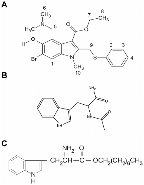
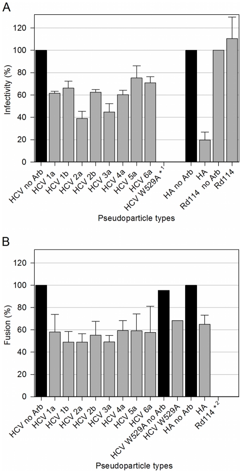
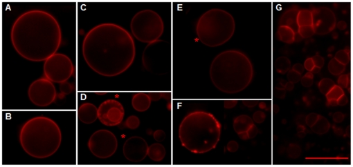
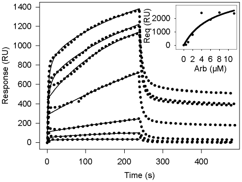
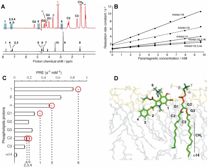
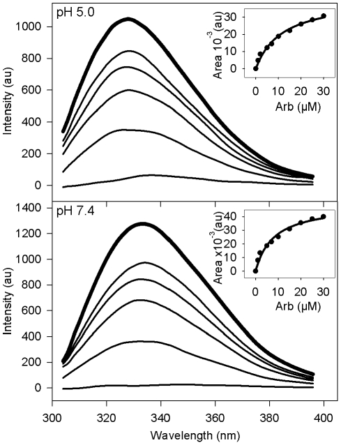
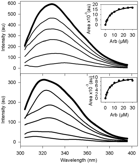
The broad-spectrum antiviral arbidol (Arb) inhibits cell entry of enveloped viruses by blocking viral fusion with host cell membrane. To better understand Arb mechanism of action, we investigated its interactions with phospholipids and membrane peptides. We demonstrate that Arb associates with phospholipids in the micromolar range. NMR reveals that Arb interacts with the polar head-group of phospholipid at the membrane interface. Fluorescence studies of interactions between Arb and either tryptophan derivatives or membrane peptides reconstituted into liposomes show that Arb interacts with tryptophan in the micromolar range. Interestingly, apparent binding affinities between lipids and tryptophan residues are comparable with those of Arb IC50 of the hepatitis C virus (HCV) membrane fusion. Since tryptophan residues of membrane proteins are known to bind preferentially at the membrane interface, these data suggest that Arb could increase the strength of virus glycoprotein's interactions with the membrane, due to a dual binding mode involving aromatic residues and phospholipids. The resulting complexation would inhibit the expected viral glycoprotein conformational changes required during the fusion process. Our findings pave the way towards the design of new drugs exhibiting Arb-like interfacial membrane binding properties to inhibit early steps of virus entry, i.e., attractive targets to combat viral infection.


Conflict of interest statement

Competing Interests: The authors have declared that no competing interests exist.

Figures

Figure 1
Figure 1. Chemical structures of arbidol (A), N-acetyl tryptophanamide (NATA) (B), and tryptophan octyl ester (TOE) (C).
Note that numbering in panel A refers to proton numbers, as identified in NMR (cf Figure 5 and Table 1).
Figure 2
Figure 2. Arb inhibition of cell entry and membrane fusion of HCVpp of various genotypes.
A, HCV entry assays using HCVpp in the absence or presence of 11.3 µM arbidol. Huh-7 cells were infected by co-incubating HCVpp of indicated genotype with or without Arb for 6 h. Infectivity was evaluated after 72 h by counting the percentage of GFP-positive cells, using a high-throughput flow cytometer (FACScalibur). The titer obtained in the absence of Arb was set to 100%, and the resulting percentages of infection in the presence of Arb were calculated. Results are the mean +/− SEM of 5 separate experiments. HApp are presented as control pseudoparticles sensitive to arbidol (cf also [10]), and Rd114pp insensitive to arbidol (cf also [12]). * 1, the mutant HCVpp W529A (cf [17]) are presented as a negative control of entry, displaying very low infectivity. B, Membrane fusion between HCVpp and R18-labeled liposomes was measured by recording the kinetics of lipid mixing by fluorescence spectroscopy (excitation and emission wavelengths were 560 and 590 nm, respectively), as described in the Materials and Methods section. Values of the last 30 s of fusion kinetics (final extent of fusion) were used to calculate the percentage of fusion in the presence of Arb, relative to fusion kinetics without Arb (100%). Results are the mean +/− SEM of 4 separate experiments. HApp and mutant HCVpp W529A were taken as controls. * 2: no fusion was observed for Rd114pp.
Figure 3
Figure 3. Arb interacts with lipid bilayers of giant unilamellar liposomes.
GUV composed of PC∶chol∶R18 (2 nmol) were electroformed in water and observed by optical epi-fluorescence microscopy (A). Various concentrations of Arb in water were added to GUV, for final Arb-to-lipid molar ratios of: B, 1∶40; C, 1∶20; D, 1∶10; E, 1∶1; F, 10∶1 and G. 20∶1. Asterisks indicate small invaginations (panel D) or occasional GUV flickering (panel E). Bar, 25 µm.
Figure 4
Figure 4. Binding of Arb to immobilized DMPC membranes.
Arb in water at concentrations of 0.5, 1, 2, 4, 6, 8 and 11.3 µM was injected over immobilized DMPC membranes (ca. 5000 resonance units) for 4 min at a flow rate of 20 µl/min, followed by water. Blank curves without Arb were substracted from those obtained with Arb. Inset, representative set of data of non-linear regression fits to the equilibrium resonance signal (Req), obtained by extrapolation to infinite time (see Materials and Methods), vs Arb concentration, used to obtain apparent equilibrium dissociation constant (KD) as well as the maximum binding capacities (Rmax). Kinetics were reproduced 4 times. Dotted curves represent the sensorgram and solid curves the non-linear fit. RU, resonance units.
Figure 5
Figure 5. NMR of Arb into lipid bicelles.
A, 1H NMR spectrum of Arb in D2O (in black) and in DMPC/DHPC bicelles (in red) with [Arb]/[lipids] = 1/15 and T = 305 K. B, influence of the concentration of the paramagnetic agent Gd(DTPA-BMA) on the proton relaxation times for Arb in the bicelle system. C, paramagnetic relaxation enhancements (PRE) measured on Arb (marked by dotted vertical lines) and on the phospholipid protons (marked by histogram bars). The phospholipid is used as a yardstick to roughly estimate the arbidol proton positions inside the membrane. Red circles indicate the yardstick marker closest to a given arbidol PRE value. Error bars indicate the standard deviation derived from the calculation of PRE. Error bars for Arbidol are comparable. D, sketch of the positioning of Arb in a DMPC membrane system. The Arb molecule was produced by generating an extended structure, and regularized by 1000 cycles of a Powell type minimization using XPLOR-NIH . The positioning in the membrane system was done manually by taking into account the relative proton depth measured by the PRE (panel C).
Figure 6
Figure 6. Indole emission fluorescence spectrum of TOE into PC∶chol liposomes.
PC∶chol (70∶30 molar ratio) liposomes containing TOE (5 µM final, lipid-to-TOE ratio 20∶1) were equilibrated to 37°C in PBS at pH 7.4 or 5.0, in the absence (bold line) or presence (standard lines) of increasing concentrations of Arb (2, 5, 10, 25 and 100 µM). Indole fluorescence was measured between 300 and 400 nm, with excitation at 286 nm. The apparent affinity of Arb toward TOE was calculated from the plot of the difference ΔA between spectral areas (AUC) of TOE without or with Arb (ΔA = AUCno Arb−AUCwith Arb) as a function of Arb concentration (see inset for a range of Arb concentrations between 0 and 30 µM) (see KD values reported in Table 3).
Figure 7
Figure 7. Trp emission fluorescence spectrum of an HCV model peptide into PC∶chol liposomes.
PC∶chol (70∶30 molar ratio) liposomes containing HCV NS4A peptide (KKGGSTWVLVGGVLAALAAYCLSTGSGGKK, 5 µM final, lipid-to-peptide ratio 20∶1) were equilibrated to 37°C in PBS at pH 7.4 or 5.0, in the absence (bold line) or presence (standard lines) of increasing concentrations of Arb (2, 5, 10, 25 and 100 µM). Trp fluorescence was measured between 300 and 400 nm, with excitation at 286 nm. The apparent affinity of Arb toward Trp was calculated from the plot of the difference ΔA between areas under the curve (AUC) of peptide without or with Arb (ΔA = AUCno Arb−AUCwith Arb) as a function of Arb concentration (see inset for a range of Arb concentrations between 0 and 30 µM) (see KD values reported in Table 4).

References

    1. Polyak SJ, Morishima C, Lohmann V, Pal S, Lee DY, et al. Identification of hepatoprotective flavonolignans from silymarin. Proc Natl Acad Sci U S A. 2010;107:5995–5999. - PMC - PubMed
    1. Wagoner J, Negash A, Kane OJ, Martinez LE, Nahmias Y, et al. Multiple effects of silymarin on the hepatitis C virus lifecycle. Hepatology. 2010;51:1912–1921. - PMC - PubMed
    1. Ahmed-Belkacem A, Ahnou N, Barbotte L, Wychowski C, Pallier C, et al. Silibinin and related compounds are direct inhibitors of hepatitis C virus RNA-dependent RNA polymerase. Gastroenterology. 2010;138:1112–1122. - PubMed
    1. Chai H, Zhao Y, Zhao C, Gong P. Synthesis and in vitro anti-hepatitis B virus activities of some ethyl 6-bromo-5-hydroxy-1H-indole-3-carboxylates. Bioorg Med Chem. 2006;14:911–917. - PubMed
    1. Brooks MJ, Sasadeusz JJ, Tannock GA. Antiviral chemotherapeutic agents against respiratory viruses: where are we now and what's in the pipeline? Curr Opin Pulm Med. 2004;10:197–203. - PubMed

Publication types

MeSH terms