. 2011 Jan 24;6(1):e16394.
doi: 10.1371/journal.pone.0016394.

The effect of a DNA repair gene on cellular invasiveness: XRCC3 over-expression in breast cancer cells

Affiliations

The effect of a DNA repair gene on cellular invasiveness: XRCC3 over-expression in breast cancer cells

Veronica L Martinez-Marignac et al. PLoS One. .

Abstract

Over-expression of DNA repair genes has been associated with resistance to radiation and DNA-damage induced by chemotherapeutic agents such as cisplatin. More recently, based on the analysis of genome expression profiling, it was proposed that over-expression of DNA repair genes enhances the invasive behaviour of tumour cells. In this study we present experimental evidence utilizing functional assays to test this hypothesis. We assessed the effect of the DNA repair proteins known as X-ray complementing protein 3 (XRCC3) and RAD51, to the invasive behavior of the MCF-7 luminal epithelial-like and BT20 basal-like triple negative human breast cancer cell lines. We report that stable or transient over-expression of XRCC3 but not RAD51 increased invasiveness in both cell lines in vitro. Moreover, XRCC3 over-expressing MCF-7 cells also showed a higher tumorigenesis in vivo and this phenotype was associated with increased activity of the metalloproteinase MMP-9 and the expression of known modulators of cell-cell adhesion and metastasis such as CD44, ID-1, DDR1 and TFF1. Our results suggest that in addition to its' role in facilitating repair of DNA damage, XRCC3 affects invasiveness of breast cancer cell lines and the expression of genes associated with cell adhesion and invasion.


Conflict of interest statement

Competing Interests: The authors have declared that no competing interests exist.

Figures

Figure 1
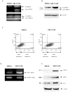
Figure 1. Assessing the invasion phenotype of XRCC3 over-expressing cells.
(A) Independent XRCC3 and RAD51 transient transfections were assayed and their protein expression was assessed through Western Blot. (B) MOCK and RAD51 or XRCC3 OE BT20 and MCF-7 cells were seeded onto Matrigel covering the surface of a porous membrane. Cells were incubated for 48 hrs to allow penetration of the matrix. Invading cells were subsequently fixed using 3.7% formaldehyde solution and stained using Hematoxylin-EosinY (Thermo Scientific). Migrating/invading cells were counted. (C) Data was then plotted as mean cells per field ±SD for a minimum of three separate experiments, each in duplicate. XRCC3 OE cells had a much higher degree of basal motility and invasion than did their MOCK BT20 and MCF-7 counterparts (2 fold higher, P<0.01) while RAD51 OE cells were not significantly different from MOCK cells.
Figure 2
Figure 2. Characterization of the expression of proteins associated with ECM metabolism and invasion.
(A) Zymography shows MMP-2 and MMP-9 activity in serum free MCF-7 conditioned medium. The gelanolytic bands of 84 (active) and 92 kDa (pro-active) MMP-9 were increased two- and more than three-fold, respectively (P = 0.045 and P<0.001), in XRCC3 OE cells with respect to MOCK cells (P<0.01); while there were no differences for MMP-2 bands. (B) MMP-9 protein levels were increased 4-fold in XRCC3 OE cells compared to MOCK cells (P = 0.033). (C) Using flow cytometry, we found that XRCC3 OE cells expressed significantly higher levels of CD44 protein than MOCK cells (two-fold, P = 0.041). (D) Using semi-quantitative reverse transcription PCR a two-fold increase in CD44 mRNA expression was observed in XRCC3 OE cells (P<0.001). (E) ID-1 and DDR1 protein levels were significantly increased (P = 0.043 and P = 0.029, respectively) in XRCC3 OE cells with respect to MOCK cells.
Figure 3
Figure 3. XRCC3 over-expression and MCF-7 xenograft growth in SCID mice.
(A) Representative photomicrographs (×20) of anti-PCNA (Novocastra) and anti-XRCC3 (Abcam, Inc.) IHC performed on XRCC3 OE and MOCK consecutive tumour sections (Left) and anti-XRCC3 expression histograms (Right). The bar graphs represent the percentage of cells with high intensity nuclear staining for anti-XRCC3 (mean +/- standard deviation). The expression of PCNA showed no significant differences between the two tumour sections, while XRCC3 expression were significantly higher in XRCC3 OE tumours (P = 0.029), * significantly different P<0.05; (B) Kaplan Meier Survival plot showing that mice injected with XRCC3 OE cells tumours earlier and in a significantly higher incidence (P = 0.028) than the MOCK cells injected mice. After 12 weeks 4/7 and 1/8 mice showed tumours for XRCC3 OE and MOCK injections, respectively.

References

    1. Jemal A, Siegel R, Ward E, Murray T, Xu J, et al. Cancer statistics, 2007. CA Cancer J Clin. 2007;57:43–66. - PubMed
    1. Toronto: Canadian Cancer Society's Steering Committee; Canadian Cancer Statistics 2010.
    1. Mimeault M, Batra SK. New promising drug targets in cancer- and metastasis-initiating cells. Drug Discov Today. 2010;15:354–364. - PMC - PubMed
    1. Aloyz R, Xu ZY, Bello V, Bergeron J, Han FY, et al. Regulation of cisplatin resistance and homologous recombinational repair by the TFIIH subunit XPD. Cancer Res. 2002;62:5457–5462. - PubMed
    1. Liu C, Zhou S, Begum S, Sidransky D, Westra WH, et al. Increased expression and activity of repair genes TDP1 and XPF in non-small cell lung cancer. Lung Cancer. 2007;55:303–311. - PMC - PubMed

Publication types

Substances