. 2011 Jan 24;6(1):e16202.
doi: 10.1371/journal.pone.0016202.

It takes two-skilled recognition of objects engages lateral areas in both hemispheres

Affiliations

It takes two-skilled recognition of objects engages lateral areas in both hemispheres

Merim Bilalić et al. PLoS One. .

Abstract

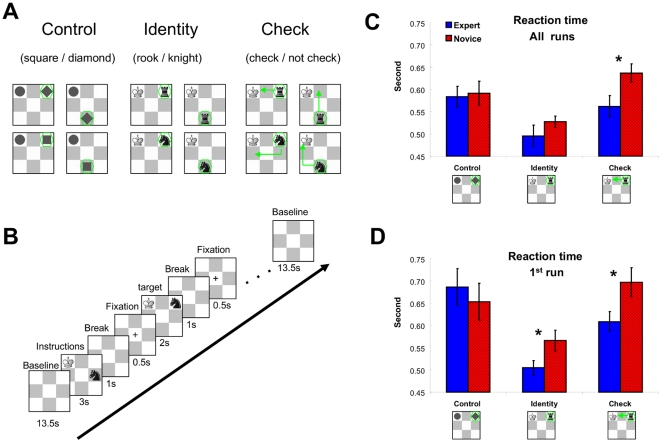
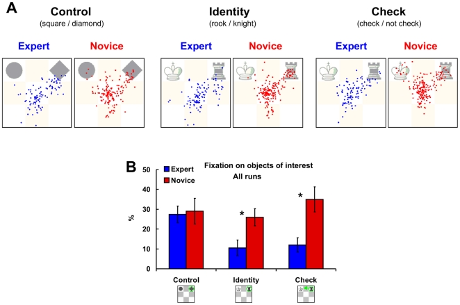
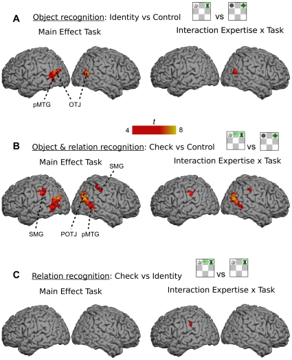
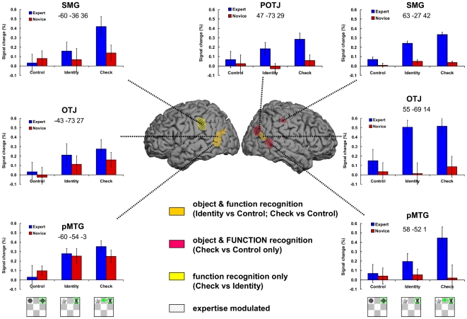
Our object recognition abilities, a direct product of our experience with objects, are fine-tuned to perfection. Left temporal and lateral areas along the dorsal, action related stream, as well as left infero-temporal areas along the ventral, object related stream are engaged in object recognition. Here we show that expertise modulates the activity of dorsal areas in the recognition of man-made objects with clearly specified functions. Expert chess players were faster than chess novices in identifying chess objects and their functional relations. Experts' advantage was domain-specific as there were no differences between groups in a control task featuring geometrical shapes. The pattern of eye movements supported the notion that experts' extensive knowledge about domain objects and their functions enabled superior recognition even when experts were not directly fixating the objects of interest. Functional magnetic resonance imaging (fMRI) related exclusively the areas along the dorsal stream to chess specific object recognition. Besides the commonly involved left temporal and parietal lateral brain areas, we found that only in experts homologous areas on the right hemisphere were also engaged in chess specific object recognition. Based on these results, we discuss whether skilled object recognition does not only involve a more efficient version of the processes found in non-skilled recognition, but also qualitatively different cognitive processes which engage additional brain areas.


Conflict of interest statement

Competing Interests: The authors have declared that no competing interests exist.

Figures

Figure 1
Figure 1. Stimuli and behavioural data.
(A) Tasks: Control task required to identify geometrical shapes (square or diamond), Identity task to indicate whether the presented piece is a rook or a knight, and Check task to indicate whether the black piece (knight or rook) gives the white king check. The four different stimulus exemplars in each condition are presented (see Methods for explanation). (B) Diagram depicting the trial structure. Each block started with an empty 3×3 board, presented for 13.5 s, which acted as a baseline. The baseline was followed by an instruction/task cue for 3 s indicating the required task. After the instruction, an empty 3×3 board appeared for 1 s and served as break. Then a black fixation cross appeared in the middle of the 3×3 board. The cross lasted for 0.5 s and was used to inform players about the upcoming stimulus. The target stimulus lasted until response or maximally for 2 s. Independent on response times, the next trial started 2 s after stimulus onset with the presentation of the 1 s break and the fixation cross. There were 4 trials in a block and after each block the baseline was presented. (C) Reaction time (RT; in seconds) averaged for experts and novices in the Control, Identity, and Check tasks over all runs (whole session). (D) Reaction time averaged for experts and novices in the Control, Identity, and Check tasks in the first run (first quarter of the session). Error bars indicate the standard error of the mean (SEM). *p<.05 in a two tailed t-test for independent samples (experts versus novices).
Figure 2
Figure 2. Eye movement data.
(A) All fixations of experts (blue dots) and novices (red dots) on an example stimulus in the Control (left), Identity (middle), and Check (right) task. (B) Average percentage of fixation that falls on objects of interests in a trail in experts and novices in the Control, Identity, and Check tasks averaged across all runs. Error bars indicate SEM. * p <.05 and †p <.10 in a two tailed t-test for independent samples (experts versus novices).
Figure 3
Figure 3. Neuroimagining data.
(A) The network of brain areas activated in recognition of chess object across all runs (whole session) – contrast Identity vs Control task (left side) and its interaction with expertise (right side). (B) The network of brain areas activated in recognition of chess object and their functions across all runs (whole session) – contrast Check vs Control task (left side) and its interaction with expertise (right side). (C) The network of brain areas activated in recognition of object functions across all runs (whole session) – contrast Check vs Identity task (left side) and its interaction with expertise (right side). The comparisons were based on p<.05 (corrected) and clusters of 5 or more voxels. The interaction between task and expertise in (A) and (C) were based on a lower threshold of p<.00001 (uncorrected). The significant areas included bilateral posterior middle temporal gyrus (pMTG), bilateral occipito-temporal junction (OTJ), right parieto-occipito-temporal junction (POTJ), and bilateral supramarginal gyrus (SMG). The MNI coordinates can be found below the labels of the ROIs in Figure 4.
Figure 4
Figure 4. Neuroimagining data summary.
Presents the object specific network based on the three comparisons in Figure 3. Orange color indicates the areas activated in both Identity vs Control, and Check vs Control task comparisons (both separately, not conjunction analsysis); Red indicates the areas activated only in the Check vs Control task (FUNCTION is in capital letters to emphasize that this area explicitly involves function of objects, unlike the areas in orange); Yellow indicates the region significantly activated in the Check vs Identity. Please note the colours are transparent and on the surface of the brain image may look slightly different. The areas modulated by expertise have additional black stripes. In each of these areas the regions of interests (ROI) were taken by selecting the voxels within 3 mm3 of the peak activation (see MNI coordinates below the ROI labels). The activation levels (percent signal change relative to baseline) were extracted for each individual player and averaged across groups and tasks for the first run only (similar results were obtained when all runs were used).

References

    1. Grill-Spector K, Malach R. The human visual cortex. Ann Rev Neurosci. 2004;27:649–677. - PubMed
    1. Lewis JW. Cortical networks related to human use of tools. Neuroscientist. 2006;12:211–231. - PubMed
    1. Noppeney U. The neural systems of tool and action semantics: a perspective from functional imaging. J Physiology. 2008;102:40–49. - PubMed
    1. Goodale MA, Milner D. Separate visual pathways for perception and action. T Neurosci. 1992;15:20–25. - PubMed
    1. Ungerleider LG, Mishkin M. Two cortical visual systems. In: Ingle MAGDJ, Mansfield RJW, editors. Analysis of Visual Behavior. Cambridge: The MIT Press; 1982. pp. 549–586.

Publication types