. 2011 Jan 20;6(1):e16398.
doi: 10.1371/journal.pone.0016398.

The Hsc/Hsp70 co-chaperone network controls antigen aggregation and presentation during maturation of professional antigen presenting cells

Affiliations

The Hsc/Hsp70 co-chaperone network controls antigen aggregation and presentation during maturation of professional antigen presenting cells

Nadja Kettern et al. PLoS One. .

Abstract

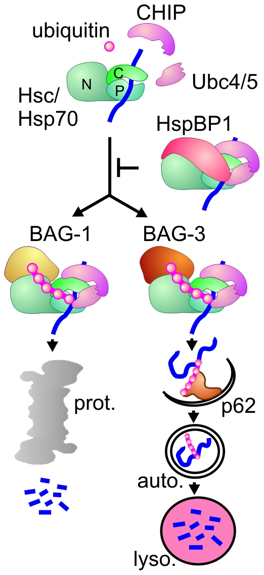
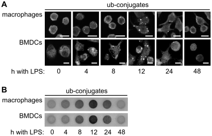
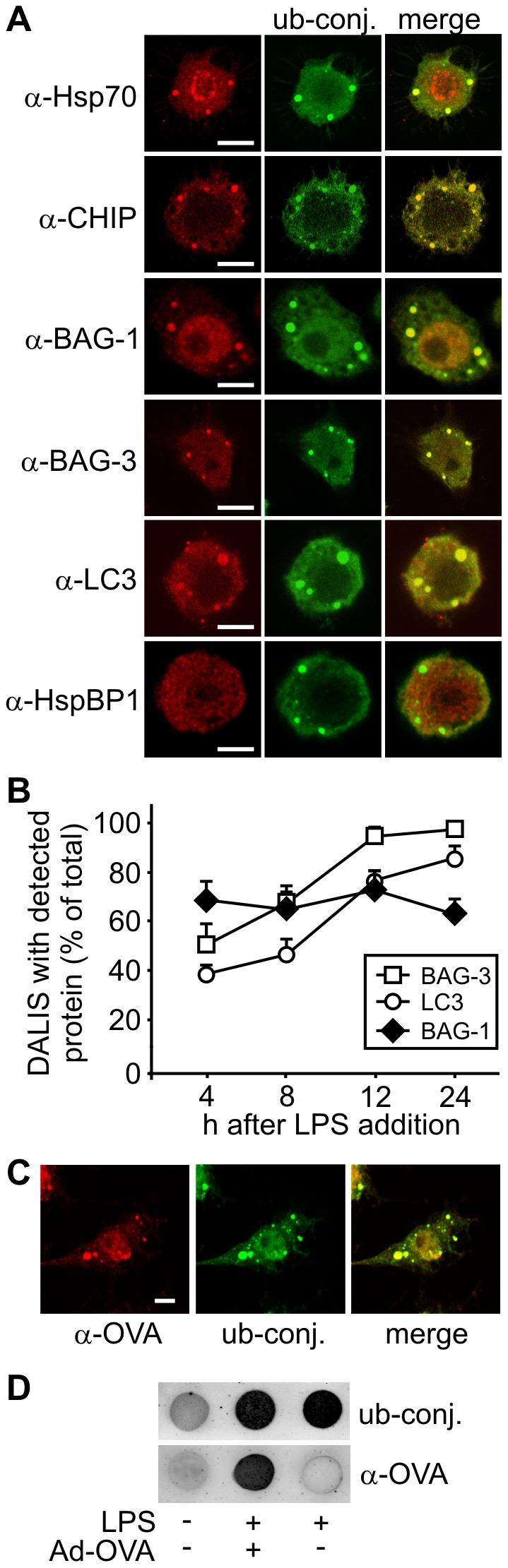
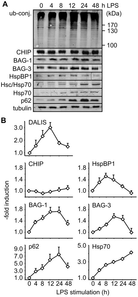
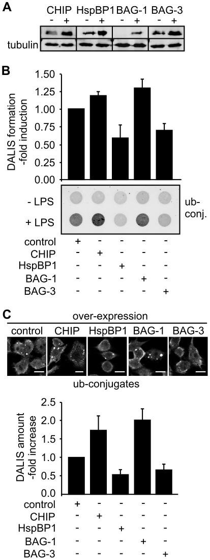
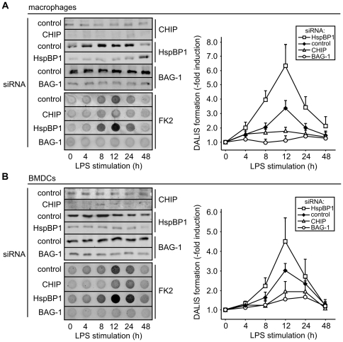
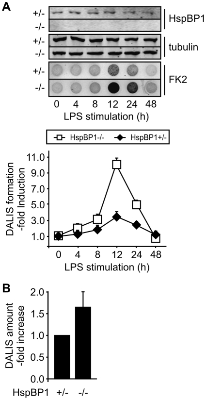
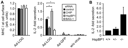
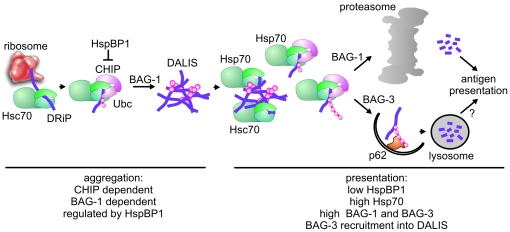
The maturation of mouse macrophages and dendritic cells involves the transient deposition of ubiquitylated proteins in the form of dendritic cell aggresome-like induced structures (DALIS). Transient DALIS formation was used here as a paradigm to study how mammalian cells influence the formation and disassembly of protein aggregates through alterations of their proteostasis machinery. Co-chaperones that modulate the interplay of Hsc70 and Hsp70 with the ubiquitin-proteasome system (UPS) and the autophagosome-lysosome pathway emerged as key regulators of this process. The chaperone-associated ubiquitin ligase CHIP and the ubiquitin-domain protein BAG-1 are essential for DALIS formation in mouse macrophages and bone-marrow derived dendritic cells (BMDCs). CHIP also cooperates with BAG-3 and the autophagic ubiquitin adaptor p62 in the clearance of DALIS through chaperone-assisted selective autophagy (CASA). On the other hand, the co-chaperone HspBP1 inhibits the activity of CHIP and thereby attenuates antigen sequestration. Through a modulation of DALIS formation CHIP, BAG-1 and HspBP1 alter MHC class I mediated antigen presentation in mouse BMDCs. Our data show that the Hsc/Hsp70 co-chaperone network controls transient protein aggregation during maturation of professional antigen presenting cells and in this way regulates the immune response. Similar mechanisms may modulate the formation of aggresomes and aggresome-like induced structures (ALIS) in other mammalian cell types.


Conflict of interest statement

Competing Interests: The authors have declared that no competing interests exist.

Figures

Figure 1
Figure 1. The co-chaperones CHIP, BAG-1, BAG-3, and HspBP1 regulate chaperone-assisted degradation.
Chaperone-assisted degradation is initiated when CHIP binds to the carboxy-terminus of Hsc/Hsp70 (‘C’). After CHIP-mediated recruitment of ubiquitin conjugating enzymes of the Ubc4/5 family, the chaperone-bound protein substrate is modified by attachment of a ubiquitin chain. The co-chaperones BAG-1, BAG-3 and HspBP1 regulate CHIP function. They are able to bind to the amino terminal ATPase domain of Hsc/Hsp70 (‘N’) at the same time when CHIP occupies the carboxy-terminus. Binding of BAG-1 initiates sorting to the proteasome (‘prot.’), whereas BAG-3 triggers the recruitment of the autophagic ubiquitin adaptor p62 and thus facilitates substrate degradation through the autophagosome-lysosome pathway. In contrast, HspBP1 inhibits the ubiquitin ligase activity of CHIP in the formed chaperone complex and thereby abrogates chaperone-assisted degradation. (‘P’ - peptide binding domain of Hsc/Hsp70; ‘auto.’ - autophagosome; ‘lyso.’ - lysosome).
Figure 2
Figure 2. DALIS are detectable by immune fluorescence and a filter trap assay.
(A) Following addition of LPS to RAW309 macrophages and mouse BMDCs ubiquitin conjugates were detected by immune fluorescence using the FK2 antibody. Transient DALIS formation peaks around 12 h after LPS addition. Scale bars: 10 µm. (B) Immune cells were lysed at the indicated time points during LPS-induced maturation in detergent containing buffer and passed through a nitrocellulose filter. Detergent insoluble ubiquitin conjugates were detected on the filter with the FK2 antibody. 50 µg of cell lysate were passed through each dot.
Figure 3
Figure 3. Diverse proteostasis components co-localize with antigens in DALIS.
(A) Using specific antibodies against diverse proteostasis components Hsp70, CHIP, BAG-1, BAG-3, and LC3 were detected in DALIS between 12 and 24 h after LPS addition to RAW309 macrophages. In contrast, the co-chaperone HspBP1 did not colocalize with ubiquitin conjugates in DALIS. DALIS were detected with the FK2 antibody that recognizes ubiquitin conjugates (‘ub-conj.’). Scale bars correspond to 5 µm. (B) The amount of DALIS that stained positive for BAG-1, BAG-3 and LC3 were quantified at the indicated time points after LPS addition. Error bars represent sem from three independent experiments. (C) RAW309 macrophages were infected with an ovalbumin expressing adenovirus and stimulated with LPS for 12 h prior to immune fluorescence. Ovalbumin was detected in DALIS with a specific antibody (‘α-OVA’), while ubiquitin conjugates (‘ub-conj.’) were visualized with the FK2 antibody. Scale bar: 5 µm. (D) RAW309 macrophages were infected with Ad-OVA and stimulated with LPS for 12 h. Control cells were left untreated or treated with LPS only. Cells were lysed in detergent containing buffer and 50 µg of total protein were passed over a nitrocellulose membrane. DALIS were detected with FK2 antibody (‘ub-conj.’) and ovalbumin with a specific antibody (‘α-OVA’).
Figure 4
Figure 4. Immune cell maturation involves significant changes of the Hsc/Hsp70 chaperone machinery.
(A) Alteration in the levels of ubiquitin conjugates (‘ub-conj.’) and proteostasis components during LPS-induced maturation of RAW309 macrophages were investigated by immune blotting with specific antibodies. (B) Expression of proteostasis components as determined in Figure 4A were quantified and correlated with DALIS formation as determined by filter trap assays (see Figure 2B). Error bars represent sem from at least three independent experiments.
Figure 5
Figure 5. Over-expression of CHIP and BAG-1 increases DALIS formation, whereas HspBP1 and BAG-3 attenuate formation.
(A) Co-chaperone levels were determined by immune blotting with specific antibodies following transfection of RAW309 macrophages with equal amounts of empty pcDNA3.1 plasmid (‘−’) or the same vector containing the coding region for the indicated co-chaperone (‘+’). Cell lysates were prepared 24 h after transfection. (B) RAW309 macrophages were transfected with empty plasmid (‘control’) or an equal amount of a co-chaperone encoding plasmid for 24 h followed by LPS addition for 12 h. DALIS formation was analyzed using the filter trap assay. Detergent insoluble ubiquitin conjugates (‘ub-conj.’) were detected on the filter with FK2 antibody and signal intensity was quantified. Value for LPS-stimulated control cells was set to 1. Error bars represent sem from at least three independent experiments. (C) Representative immune fluorescence micrographs of transfected macrophages after over-expression of the indicated co-chaperones for 24 h followed by LPS stimulation for 12 h. Lower panel shows a quantification of obtained data. Number of DALIS observed in control cells were set to 1. Error bars represent sem from at least three independent experiments. Scale bars: 10 µm.
Figure 6
Figure 6. CHIP and BAG-1 are essential for DALIS formation, whereas HspBP1 exerts an attenuating function.
(A) Co-chaperones were depleted from RAW309 macrophages by addition of specific siRNAs for 24 h followed by addition of LPS. Control cells received an equal amount of scrambled siRNA. At indicated time points cell extracts were prepared and analyzed by immune blotting or subjected to a filter trap assay for DALIS detection. Signal intensity for detergent insoluble ubiquitin conjugates was quantified. Value at time point zero for control cells was set to 1. Error bars represent sem from three independent experiments. (B) Same as under A, except for the use of mouse BMDCs.
Figure 7
Figure 7. BMDCs isolated from hspBP1 −/− mice show increased DALIS formation during maturation.
(A) BMDCs were isolated from hspBP1 −/− mice and heterozygous siblings and treated with LPS. At indicated time points cell extracts were prepared and analyzed by immune blotting or subjected to the filter trap assay for DALIS detection. Signal intensity for detergent insoluble ubiquitin conjugates was quantified. Value at time point zero for heterozygous cells was set to 1. Error bars represent sem from three independent experiments. (B) DALIS were quantified in BMDCs from hspBP1−/− mice and heterozygous siblings by immune fluorescence with FK2 antibody after LPS treatment for 12 h. Error bars represent sem from three independent experiments.
Figure 8
Figure 8. Hsc/Hsp70 co-chaperones regulate MHC class I mediated antigen presentation.
(A) After siRNA mediated depletion of the indicated co-chaperones BMDCs were infected with Ad-LOG and stimulated with LPS. After 12 h cell surface localized MHC class I molecules were quantified by FACS analysis (left panel). Fluorescence of control cells, which received a scrambled siRNA, was set to 1. Error bars represent sem from three independent experiments. To monitor antigen presentation cells treated under the same conditions were washed and ovalbumin-specific CD8+ T cells (OT-1) were added. Co-cultivation was performed for 24 h. Presentation on MHC class I complexes was quantified by determining interleukin-2 (IL-2) production of OT-1 cells by ELISA (right panel). Values for control cells receiving a scrambled siRNA were set to 1. Error bars represent sem of at least three independent experiments. Asterisks indicate significance as determined by the two-tailed Student's t-test: control/CHIP: *P = 0.018; control/HspBP1: *P = 0.030; control/BAG-1: *P = 0.011 (B) Wild-type, heterozygous hspBP1 +/−, and homozygous hspBP1 −/− mice received an intravenous injection of recombinant influenza A PR/8/43 expressing the SIINFEKL peptide. Spleen cells were isolated after 12 h und incubated with OT-1 cells to monitor antigen presentation on MHC class I molecules based on IL-2 production. Values for wild-type mice were set to 1. Error bars represent sem of three independent experiments.
Figure 9
Figure 9. The Hsc/Hsp70 co-chaperone network controls DALIS formation during immune cell maturation.
Immune cell maturation can be separated in two distinct phases that are characterized by distinct chaperone environments. During early stages of immune cell maturation, characterized by DALIS formation (aggregation phase), Hsc/Hsp70 binds defective ribosomal products (DRiPs) following their translation. Recruitment of CHIP leads to the ubiquitylation of DRiPs, which provides a sorting signal for sequestration into DALIS. Ubiquitylation activity is regulated by the CHIP-inhibitor HspBP1. DALIS formation also requires BAG-1. Late stages of maturation (presentation phase), which involve antigen presentation and clearance of DALIS, are characterized by decreased HspBP1 levels and induction of Hsp70, BAG-1, and BAG-3. These changes stimulate DRiP processing and presentation. In addition, BAG-3 is recruited to DALIS to facilitate aggregate clearance by chaperone-assisted selective autophagy in cooperation with p62. It remains to be seen whether this autophagy pathway contributes to MHC class I mediated presentation (‘?’).

References

    1. Balch WE, Morimoto RI, Dillin A, Kelly JW. Adapting proteostasis for disease intervention. Science. 2008;319:916–919. - PubMed
    1. Finkbeiner S, Cuervo AM, Morimoto RI, Muchowski PJ. Disease-modifying pathways in neurodegeneration. J Neurosci. 2006;26:10349–10357. - PMC - PubMed
    1. Brodsky JL, Chiosis G. Hsp70 molecular chaperones: emerging roles in human disease and identification of small molecule modulators. Curr Top Med Chem. 2006;6:1215–1225. - PubMed
    1. Hartl FU, Hayer-Hartl M. Converging concepts of protein folding in vitro and in vivo. Nat Struct Mol Biol. 2009;16:574–581. - PubMed
    1. Kettern N, Dreiseidler M, Tawo R, Höhfeld J. Chaperone-assisted degradation: multiple paths to destruction. Biol Chem. 2010;391:481–489. - PubMed

Publication types