. 2011 Jan 20;6(1):e16401.
doi: 10.1371/journal.pone.0016401.

The CCCTC-binding factor (CTCF) of Drosophila contributes to the regulation of the ribosomal DNA and nucleolar stability

Affiliations

The CCCTC-binding factor (CTCF) of Drosophila contributes to the regulation of the ribosomal DNA and nucleolar stability

Paola A Guerrero et al. PLoS One. .

Abstract

In the repeat array of ribosomal DNA (rDNA), only about half of the genes are actively transcribed while the others are silenced. In arthropods, transposable elements interrupt a subset of genes, often inactivating transcription of those genes. Little is known about the establishment or separation of juxtaposed active and inactive chromatin domains, or preferential inactivation of transposable element interrupted genes, despite identity in promoter sequences. CTCF is a sequence-specific DNA binding protein which is thought to act as a transcriptional repressor, block enhancer-promoter communication, and delimit juxtaposed domains of active and inactive chromatin; one or more of these activities might contribute to the regulation of this repeated gene cluster. In support of this hypothesis, we show that the Drosophila nucleolus contains CTCF, which is bound to transposable element sequences within the rDNA. Reduction in CTCF gene activity results in nucleolar fragmentation and reduced rDNA silencing, as does disruption of poly-ADP-ribosylation thought to be necessary for CTCF nucleolar localization. Our data establish a role for CTCF as a component necessary for proper control of transposable element-laden rDNA transcription and nucleolar stability.


Conflict of interest statement

Competing Interests: The authors have declared that no competing interests exist.

Figures

Figure 1
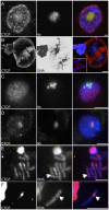
Figure 1. Indirect immunofluorescence reveals CTCF as a component of the nucleoli of Drosophila cells.
(A) Confocal microscopy of whole mount third instar larval salivary gland nucleus. CTCF and fibrillarin (fib) shown separately, and merged with DNA (blue). (B) Higher magnification of nucleolus showing CTCF, DNA, and color merge. The DNA-only separation has been inverted and non-linearly adjusted for bright and contrast to reveal the filamentous structure of the DNA within the nucleolus. Inset in merged image shows a different nucleus with the CTCF-containing nucleolus in the context of CTCF-banded chromosome arms. (C) Epifluorescence microscopy of S2 tissue culture cell nucleus. CTCF and fibrillarin shown separately, and merged with DNA. (D) Confocal microscopy of third instar larval diploid interphase neuroblast nucleus. CTCF and fibrillarin shown separately, and merged with DNA (blue). (E) Epifluorescence microscopy of condensed mitotic X chromosome (arrow points to rDNA locus) from a third instar larval diploid metaphase neuroblast. The panoply of CTCF sites in the euchromatin are visible as immunofluorescence on the chromosome arms. (F) Epifluorescence microscopy of condensed mitotic Y chromosome from a third instar larval metaphase diploid neuroblast. For (E) and (F), CTCF is shown separately from a color merge with DNA. Scale bars 5 µm.
Figure 2
Figure 2. Chromatin Immunoprecipitation of CTCF identifies binding to the rDNA locus.
(A) Map showing structure of typical rDNA repeat unit. NTS  =  non-transcribed spacer, ETS  =  external transcribed spacer, ITS  =  internal transcribed spacers, 18S, 5.8S, 2S, and 28S are final rRNA products, R1 and R2 are transposable element insertions (dotted lines indicate insertion sites in the 28S). Numbers indicate location of potential or predicted CTCF binding sites - all sites are shown, indicated either by numbers or by vertical hash marks. Asterisks indicate location of near-consensus sites within R1 and R2, collectively used as an “out-group.” Blow-out shows detail around R1 and R2 insertion sites in the 28S sequence; primers “a”–“f” are used for R1-, R2-, and uninserted 35S specific transcript detection. (B) Real-Time PCR quantification of amplification using DNA purified from chromatin immunoprecipitated by anti-CTCF antibodies. Data are presented as average boxed by pooled standard deviations of triplicate samples from three independent experiments. White data are from sites that do not match Drosophila CTCF consensus, gray data (18, 21, 28–31) match the Drosophila consensus. All data are normalized to the pooled average of the outgroup data (*), which was then defined as 100% (dashed line). Sequences show CTCF consensus sites (18, 21, 28–31).
Figure 3
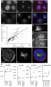
Figure 3. RNAi-mediated or mutational reduction of CTCF gene activity disrupts nucleolar structure and increases rDNA expression.
(A) Indirect immunofluorescence detection of CTCF in S2 cell culture nuclei. (B) CTCF immunodetection after three-day treatment of double-stranded RNA directed at CTCF. Images from (A) and (B) are presented with the same exposure/bright/contrast conditions. (C) Quantification of all data from untreated (circles) and double-stranded RNA treated (crosses) cells. X-axis shows CTCF intensity (fluorescence per unit time, corrected to DNA), y-axis shows fibrillarin intensity, and regression lines are for separate datasets. (D) Higher magnification of fibrillarin-containing nucleolus from control cell treated with double-stranded RNA directed at LacZ. (E) Higher magnification of fibrillarin-containing nucleolus from cell treated with double-stranded RNA directed at CTCF. (F) As in (E), but a more pronounced nucleolar vesiculation/disruption phenotype. (G) Salivary gland nuclei derived from third instar larvae mutant for CTCF. In images from (A), (B), and (G), CTCF and fibrillarin (fib) are shown separately, and merged with DNA (blue). (H) In S2 cell culture, no double-stranded RNA treatment, or treatment with double-stranded RNAs directed at LacZ have no effect on 28S rRNA (Figure 2, primers a–d), R1 (Figure 2, primers b–c) or R2 (Figure 2, primers e-f) mRNA levels, or pre-rRNA unprocessed junctions (2S-ITS2 and ITS2-28S) but treatment with double-stranded RNAs directed at CTCF increases 28S, R1, R2, and pre-RNA junction RNA species. Whole intact animals bearing homozygous mutation of CTCF 35.2 results in increased 28S, R1, R2, and pre-RNA junction RNA species compared to heterozygous CTCF 35.2/+ controls. Note discontinuity in ordinate for final R2 datum (end-hashed dashed line). All data are normalized (100%, dashed lines) to untreated cells (“no dsRNA”) and heterozygous animals (“CTCT/+”). Scale bars 5 µm.
Figure 4
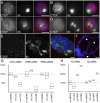
Figure 4. Disruption of poly-ADP-ribosylation decreases nucleolar CTCF, disrupts nucleolar structure, and increases rDNA expression.
(A) Indirect immunofluorescence detection of CTCF and fibrillarin (fib) in S2 cell culture, and (B) after treatment with double stranded RNA directed at LacZ. (C) Structure of nucleoli after treatment with control double-stranded RNA directed at Poly-ADP-Ribose Polymerase (PARP), or (D) Poly-ADP-Ribose Glycohydrolase (PARG). (E) Confocal microscopy of whole mount third instar larval salivary gland nuclei derived from PARG mutants. In all preceding images, CTCF and fibrillarin (fib) are shown separately, and merged with DNA (blue). (F) Squashed chromosomes from whole mount third instar larval salivary gland nuclei show retention of CTCF at euchromatic bands but loss from the nucleolus (arrowheads). (G) Double-stranded RNA directed at CTCF, PARP, or PARG and their effects on mRNA level. (H) R1 and R2 mRNA expression after treatment with double-stranded RNAs directed at LacZ (control), PARP, and PARG. For (G) and (H), data are normalized to untreated cells (100%, dashed lines). Scale bars 5 µm.
Figure 5
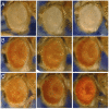
Figure 5. Maternal heterozygosity for CTCF suppresses rDNA-induced position effect variegation.
(A) Expression of white+ from P-element B486 in wild-type flies derived from wild-type (left), CTCF 35.2/+ (middle), and Df(3L)0463/+ (right) mothers. (B) Expression of white+ from P-element ROMA in wild-type flies derived from wild-type (left), CTCF 35.2/+ (middle), and Df(3L)0463/+ (right) mothers. (C) Expression of white+ from rDNA-inserted P-element D285 in wild-type flies derived from wild-type (left), CTCF 35.2/+ (middle), and Df(3L)0463/+ (right) mothers.

Similar articles

Cited by

References

    1. McStay B, Grummt I. The epigenetics of rRNA genes: from molecular to chromosome biology. Annu Rev Cell Dev Biol. 2008;24:131–157. - PubMed
    1. Miller OL, Jr, Beatty BR. Visualization of nucleolar genes. Science. 1969;164:955–957. - PubMed
    1. Percipalle P, Fomproix N, Cavellan E, Voit R, Reimer G, et al. The chromatin remodelling complex WSTF-SNF2h interacts with nuclear myosin 1 and has a role in RNA polymerase I transcription. EMBO Rep. 2006;7:525–530. - PMC - PubMed
    1. Langst G, Becker PB, Grummt I. TTF-I determines the chromatin architecture of the active rDNA promoter. EMBO J. 1998;17:3135–3145. - PMC - PubMed
    1. Huang J, Moazed D. Association of the RENT complex with nontranscribed and coding regions of rDNA and a regional requirement for the replication fork block protein Fob1 in rDNA silencing. Genes Dev. 2003;17:2162–2176. - PMC - PubMed

Publication types