. 2011 Jan 20;6(1):e16175.
doi: 10.1371/journal.pone.0016175.

Chronic respiratory aeroallergen exposure in mice induces epithelial-mesenchymal transition in the large airways

Affiliations

Chronic respiratory aeroallergen exposure in mice induces epithelial-mesenchymal transition in the large airways

Jill R Johnson et al. PLoS One. .

Abstract

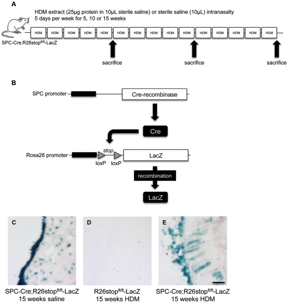
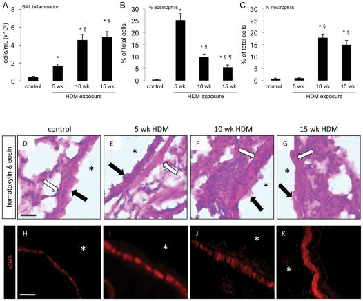
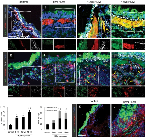
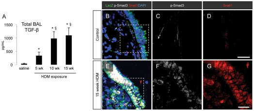
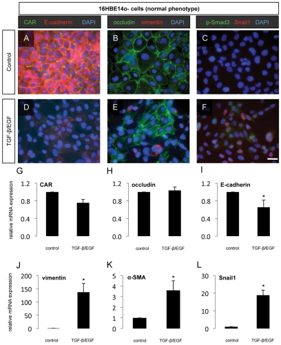
Chronic allergic asthma is characterized by Th2-polarized inflammation and leads to airway remodeling and fibrosis but the mechanisms involved are not clear. To determine whether epithelial-mesenchymal transition contributes to airway remodeling in asthma, we induced allergic airway inflammation in mice by intranasal administration of house dust mite (HDM) extract for up to 15 consecutive weeks. We report that respiratory exposure to HDM led to significant airway inflammation and thickening of the smooth muscle layer in the wall of the large airways. Transforming growth factor beta-1 (TGF-β1) levels increased in mouse airways while epithelial cells lost expression of E-cadherin and occludin and gained expression of the mesenchymal proteins vimentin, alpha-smooth muscle actin (α-SMA) and pro-collagen I. We also observed increased expression and nuclear translocation of Snail1, a transcriptional repressor of E-cadherin and a potent inducer of EMT, in the airway epithelial cells of HDM-exposed mice. Furthermore, fate-mapping studies revealed migration of airway epithelial cells into the sub-epithelial regions of the airway wall. These results show the contribution of EMT to airway remodeling in chronic asthma-like inflammation and suggest that Th2-polarized airway inflammation can trigger invasion of epithelial cells into the subepithelial regions of the airway wall where they contribute to fibrosis, demonstrating a previously unknown plasticity of the airway epithelium in allergic airway disease.


Conflict of interest statement

Competing Interests: The authors have declared that no competing interests exist.

Figures

Figure 1
Figure 1. LacZ expression pattern in SPC-Cre;R26stopfl/fl-LacZ mice.
(A) Model of chronic respiratory house dust mite (HDM) extract exposure. Mice were administered sterile saline or 25 µg of HDM extract in a volume of 10 µL 5 days a week for up to 15 consecutive weeks. Mice were sacrificed after 5, 10 or 15 weeks of HDM exposure. (B) Characterization of the reporter mice used in this study. Cre is expressed by the SPC promoter and removes the floxed stop sequence in front of the gene for LacZ under the control of the ROSA26 promoter. Thus, all lung epithelial cells stably and irreversibly express LacZ. (C–E) Enzymatic staining for β-galactosidase activity was performed on 15 µm-thick lung sections from (C) SPC-Cre;R26stopfl/fl-LacZ mice exposed to saline, (D) R26stopfl/fl-LacZ mice exposed to HDM and (E) SPC-Cre;R26stopfl/fl-LacZ exposed to HDM. Scale bar 10 µm.
Figure 2
Figure 2. Prolonged respiratory HDM exposure induces inflammation.
Mice were administered sterile saline or 25 µg of HDM extract in a volume of 10 µL 5 days a week for 5, 10 or 15 consecutive weeks. (A–C) Bronchoalveolar lavage (BAL) analysis was performed to determine total inflammatory cell infiltrate (A) and to differentiate between eosinophils (B) and neutrophils (C). * p<0.05 compared to saline control animals, § p<0.05 compared to mice exposed to HDM for 5 weeks and ¶ p<0.05 compared to mice exposed to HDM for 10 weeks. Data represent mean ± SEM, n = 10–15 mice per group from two independent experiments. (D–G) Chemical staining for hematoxylin and eosin was performed on 5 µm-thick lung sections from (D) control mice exposed to saline, (E) mice exposed to HDM for 5 weeks, (F) 10 weeks or (G) 15 weeks. * indicate airway lumen, closed arrows indicate the epithelium, open arrows indicate airway smooth muscle. (H–K) Immunofluorescent staining for α-smooth muscle actin (α-SMA) was performed on 15 µm-thick lung sections from (H) control mice exposed to saline, (I) mice exposed to HDM for 5 weeks, (J) 10 weeks or (K) 15 weeks. * indicate airway lumen. Scale bar 10 µm.
Figure 3
Figure 3. Prolonged respiratory HDM exposure induces epithelial-to-mesenchymal transition.
Lung sections (15 µm thick) were prepared from control mice and mice exposed to HDM for 5, 10 or 15 weeks and immunofluorescent staining for the co-expression of the LacZ reporter in airway epithelial cells with α-SMA and occludin (A–D) and with vimentin and E-cadherin (E–H) was performed. Scale bars 10 µm. Quantification of lung fibrosis was performed by morphometric analysis of lung sections stained for α-SMA (I) or LacZ and vimentin (J). * p<0.05 compared to saline control animals, § p<0.05 compared to mice exposed to HDM for 5 weeks. Data represent mean ± SEM, n = 5 mice per group. Additional lung sections were stained to detect procollagen I-producing cells in the airway wall in control mice and in mice exposed to HDM for 10 weeks (K, L).
Figure 4
Figure 4. Activation of TGF-β signaling pathways following chronic HDM exposure.
(A) Analysis of TGF-β levels in mouse bronchoalveolar lavage (BAL) fluid in saline controls and mice exposed to HDM for 5, 10 or 15 weeks. BAL fluid was collected at the time of sacrifice and analyzed by ELISA for the expression of mouse TGF-β1. * p<0.05 compared to saline control animals, § p<0.05 compared to mice exposed to HDM for 5 weeks. Data represent mean ± SEM, n = 8 per group from two independent experiments. (B–G) Immunofluorescent staining for the expression of LacZ, p-Smad3 and Snail1 in lung sections from control mice and mice exposed to HDM for 15 weeks.
Figure 5
Figure 5. Partial induction of EMT in 16HBE14o- cells.
(A–I) Progression through EMT was evaluated in the phenotypically normal human lung epithelial cell line 16HBE14o- 72 h after the addition of TGF-β1 (10 ng/mL) and EGF (50 ng/mL) to the culture medium. The epithelial junction proteins CAR and E-cadherin (A, D), the tight junction protein occludin and the mesenchymal marker vimentin (B, E) as well as the EMT-associated transcription factors pSmad3 and Snail1 (C, F) were assessed by immunofluorescent staining. Scale bar 10 µm. Quantification of the relative mRNA expression of CAR (G), occludin (H), E-cadherin (I), vimentin (J), α-SMA (K) and Snail1 (L) was assessed by qPCR. * p<0.05 compared to untreated cells. Data represent mean ± SEM, n = 6 from two independent experiments.
Figure 6
Figure 6. Induction of EMT in A549 cells.
Progression through EMT was evaluated in the adenocarcinoma-derived human lung epithelial cell line A549 72h after the addition of TGF-β1 (10 ng/mL) and EGF (50 ng/mL) to the culture medium. The epithelial junction proteins CAR and E-cadherin (A, D), the tight junction protein occludin and the mesenchymal marker vimentin (B, E) as well as the EMT-associated transcription factors pSmad3 and Snail1 (C, F) were assessed by immunofluorescent staining. Scale bar 10 µm. Quantification of the relative mRNA expression of CAR (G), occludin (H), E-cadherin (I), vimentin (J) ), α-SMA (K) and Snail1 (L) was assessed by qPCR.* p<0.05 compared to untreated cells. Data represent mean ± SEM, n = 6 from two independent experiments.

References

    1. Holgate ST. Pathogenesis of asthma. Clin Exp Allergy. 2008;38:872–897. - PubMed
    1. Johnson JR, Swirski FK, Gajewska BU, Wiley RE, Fattouh R, et al. Divergent immune responses to house dust mite lead to distinct structural-functional phenotypes. Am J Physiol Lung Cell Mol Physiol. 2007;293:L730–739. - PubMed
    1. Holgate ST. The airway epithelium is central to the pathogenesis of asthma. Allergol Int. 2008;57:1–10. - PubMed
    1. Kalluri R, Weinberg RA. The basics of epithelial-mesenchymal transition. J Clin Invest. 2009;119:1420–1428. - PMC - PubMed
    1. Thiery JP, Acloque H, Huang RY, Nieto MA. Epithelial-mesenchymal transitions in development and disease. Cell. 2009;139:871–890. - PubMed

Publication types