. 2011 Jan 19;6(1):e16344.
doi: 10.1371/journal.pone.0016344.

Genomewide analyses define different modes of transcriptional regulation by peroxisome proliferator-activated receptor-β/δ (PPARβ/δ)

Affiliations

Genomewide analyses define different modes of transcriptional regulation by peroxisome proliferator-activated receptor-β/δ (PPARβ/δ)

Till Adhikary et al. PLoS One. .

Abstract

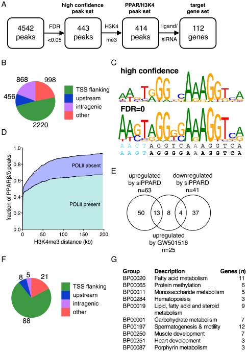
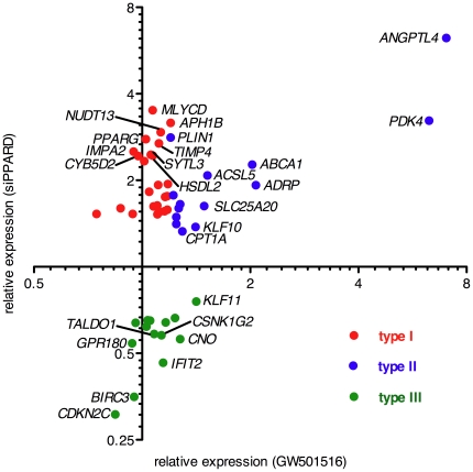
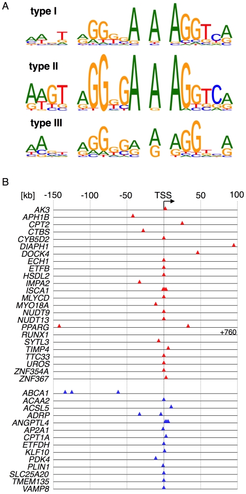
Peroxisome proliferator-activated receptors (PPARs) are nuclear receptors with essential functions in lipid, glucose and energy homeostasis, cell differentiation, inflammation and metabolic disorders, and represent important drug targets. PPARs heterodimerize with retinoid X receptors (RXRs) and can form transcriptional activator or repressor complexes at specific DNA elements (PPREs). It is believed that the decision between repression and activation is generally governed by a ligand-mediated switch. We have performed genomewide analyses of agonist-treated and PPARβ/δ-depleted human myofibroblasts to test this hypothesis and to identify global principles of PPARβ/δ-mediated gene regulation. Chromatin immunoprecipitation sequencing (ChIP-Seq) of PPARβ/δ, H3K4me3 and RNA polymerase II enrichment sites combined with transcriptional profiling enabled the definition of 112 bona fide PPARβ/δ target genes showing either of three distinct types of transcriptional response: (I) ligand-independent repression by PPARβ/δ; (II) ligand-induced activation and/or derepression by PPARβ/δ; and (III) ligand-independent activation by PPARβ/δ. These data identify PPRE-mediated repression as a major mechanism of transcriptional regulation by PPARβ/δ, but, unexpectedly, also show that only a subset of repressed genes are activated by a ligand-mediated switch. Our results also suggest that the type of transcriptional response by a given target gene is connected to the structure of its associated PPRE(s) and the biological function of its encoded protein. These observations have important implications for understanding the regulatory PPAR network and PPARβ/δ ligand-based drugs.


Conflict of interest statement

Competing Interests: The authors have declared that no competing interests exist.

Figures

Figure 1
Figure 1. Genomewide identification of PPARβ/δ binding sites in WPMY-1 cells by ChIP-Seq.
(A) PPARβ/δ enrichment at the genomic ANGPTL4 locus determined by ChIP-Seq. (B) ChIP-qPCR analysis of PPARβ/δ and RXRα binding at 12 genomic loci identified by ChIP-Seq. (C) PPARβ/δ, H3K4me3 and RNA polymerase II enrichment peaks detected by ChIP-Seq at the HSDL2 locus.
Figure 2
Figure 2. Genomewide identification of PPARβ/δ target genes by combining ChIP-Seq and transcriptional profiling.
(A) Flow chart showing consecutive steps of bioinformatic analysis for the definition of high confidence PPARβ/δ-regulated genes (target gene set). These genes are characterized by one or more peaks with an FDR<0.05 that is/are located within 200 kb of a database-defined gene (Ensembl release 58), a cluster of H3K4me3 marks, RNA polymerase II enrichment and transcriptional responsiveness to PPARD siRNA and/or the agonist GW501516. (B) Distribution of genomewide PPARβ/δ binding for all 4,542 peaks identified by ChIP-Seq. TSS flanking is defined as regions from −5000 bp to the 3′ end of the first intron, upstream regions are located within −25 kb of a transcriptional start site (TSS). (C) Consensus sequence identified by de novo motif search (MEME) of ChIP-Seq in high confidence (top) and FDR = 0 peaks (bottom). The line beneath shows the published consensus sequence ; the bottom line (boldface) shows the refined sequence derived from the present study. (D) Overlap between high confidence PPARβ/δ peaks, H3K4me3 marks and RNA polymerase II enrichment detected by parallel ChIP-Seq experiments. (E) Venn diagram showing the overlap between genes regulated by PPARD siRNA or activated by GW501516. (F) Analysis of PPARβ/δ peak distribution for the target gene set. (G) Panther biological process (BP) classification of the target gene set. The 10 most enriched BP terms describing biological processes affected by PPARβ/δ are shown.
Figure 3
Figure 3. Identification of different types of transcriptional responses to PPARβ/δ depletion and ligands.
(A–C) Differential responses to GW501516 and PPARD siRNA of PPARβ/δ target genes, classified as type I (A), type II (B) and type III (C) responses. WPMY-1 cells were treated as indicated and analyzed as in Figure 1. Data from four biological replicates are shown. Individual data points represent the average of 3 technical replicates. Horizontal lines indicate the median of 4 biological replicates. (D) PPAR subtype-specific repression of the ANGPTL4 gene by PPARβ/δ. WPMY-1 cells were transfected with PPARA, PPARG or PPARD siRNA pools, or a combination of all three pools (triple knock-down), and relative ANGPTL4 mRNA levels were measured by RT-qPCR. The efficiencies and subtype specificities of the siRNA pools are shown in Figure S4. (E) Effect of PPARD siRNA treatment on PPARβ/δ recruitment to the IMPA2, MLYCD, ANGPTL4, SLC25A20, BIRC3 and GPR180 genes.
Figure 4
Figure 4. Correlation of PPARD siRNA and GW501516 mediated gene regulation for the validated gene set.
Red: type I response, upregulated (≥1.5-fold) by PPARD siRNA and unaffected by GW501516; blue: type II response, upregulated by PPARD siRNA and induced by GW501516 (≥1.2-fold); green: type III response, down-regulated (≥1.5-fold) by PPARD siRNA. Each data point represents the average of 4 biological replicates as in Figure 3A–C.
Figure 5
Figure 5. Structural features of PPARβ/δ target genes showing type I or type II responses.
(A) Consensus PPRE motifs in ChIP-Seq peak areas of validated type I, II and III response genes derived by best-fit alignment of peak sequences with the FDR = 0 motif in Fig. 2C. (B) Locations of PPREs in PPARβ/δ enrichment peaks (type I: red; type II: blue; numbers relative to the TSS). All sites downstream of the TSS are intragenic.

References

    1. Desvergne B, Michalik L, Wahli W. Transcriptional regulation of metabolism. Physiol Rev. 2006;86:465–514. - PubMed
    1. Kilgore KS, Billin AN. PPARbeta/delta ligands as modulators of the inflammatory response. Curr Opin Investig Drugs. 2008;9:463–469. - PubMed
    1. Peters JM, Gonzalez FJ. Sorting out the functional role(s) of peroxisome proliferator-activated receptor-beta/delta (PPARbeta/delta) in cell proliferation and cancer. Biochim Biophys Acta. 2009;1796:230–241. - PMC - PubMed
    1. Grimaldi PA. Metabolic and nonmetabolic regulatory functions of peroxisome proliferator-activated receptor beta. Curr Opin Lipidol. 2010;21:186–191. - PubMed
    1. Peraza MA, Burdick AD, Marin HE, Gonzalez FJ, Peters JM. The toxicology of ligands for peroxisome proliferator-activated receptors (PPAR). Toxicol Sci. 2006;90:269–295. - PubMed

Publication types