. 2011 Jan 4;9(1):e1000568.
doi: 10.1371/journal.pbio.1000568.

The gene regulatory cascade linking proneural specification with differentiation in Drosophila sensory neurons

Affiliations

The gene regulatory cascade linking proneural specification with differentiation in Drosophila sensory neurons

Sebastián Cachero et al. PLoS Biol. .

Abstract

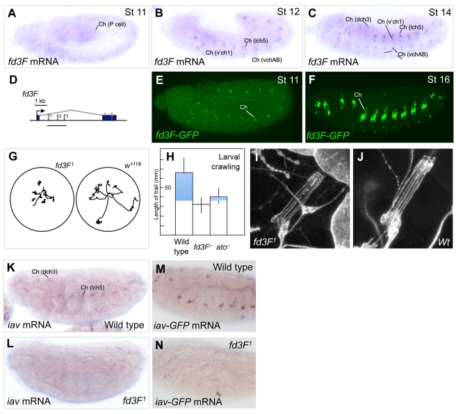
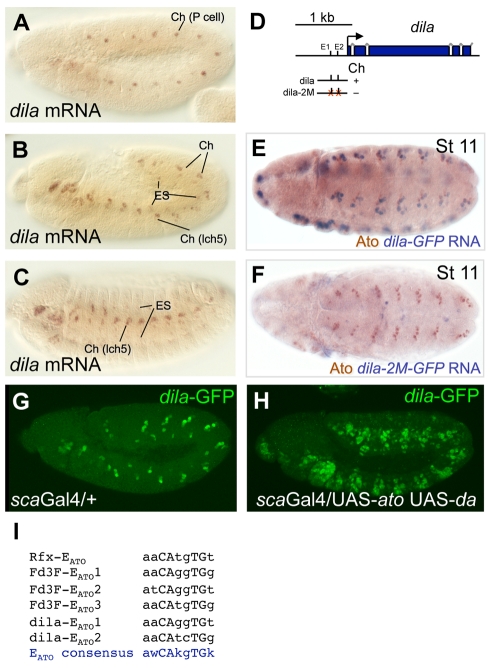
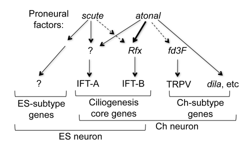
In neurogenesis, neural cell fate specification is generally triggered by proneural transcription factors. Whilst the role of proneural factors in fate specification is well studied, the link between neural specification and the cellular pathways that ultimately must be activated to construct specialised neurons is usually obscure. High-resolution temporal profiling of gene expression reveals the events downstream of atonal proneural gene function during the development of Drosophila chordotonal (mechanosensory) neurons. Among other findings, this reveals the onset of expression of genes required for construction of the ciliary dendrite, a key specialisation of mechanosensory neurons. We determine that atonal activates this cellular differentiation pathway in several ways. Firstly, atonal directly regulates Rfx, a well-known highly conserved ciliogenesis transcriptional regulator. Unexpectedly, differences in Rfx regulation by proneural factors may underlie variations in ciliary dendrite specialisation in different sensory neuronal lineages. In contrast, fd3F encodes a novel forkhead family transcription factor that is exclusively expressed in differentiating chordotonal neurons. fd3F regulates genes required for specialized aspects of chordotonal dendrite physiology. In addition to these intermediate transcriptional regulators, we show that atonal directly regulates a novel gene, dilatory, that is directly associated with ciliogenesis during neuronal differentiation. Our analysis demonstrates how early cell fate specification factors can regulate structural and physiological differentiation of neuronal cell types. It also suggests a model for how subtype differentiation in different neuronal lineages may be regulated by different proneural factors. In addition, it provides a paradigm for how transcriptional regulation may modulate the ciliogenesis pathway to give rise to structurally and functionally specialised ciliary dendrites.


Conflict of interest statement

The authors have declared that no competing interests exist.

Figures

Figure 1
Figure 1. Gene expression profiling of Ch cells.
(A) Schematic of structural features of Ch and ES organs. (B) Group of five Ch neurons in the larval lateral body wall, labelled with anti-HRP, which detects the cell body and inner dendritic segment. The approximate location of the basal body is indicated. (C) Schematic of cell lineage leading from an SOP to a Ch organ (same colour scheme as in A). Ato is expressed at the SOP stage. The time points sampled for analysis are indicated approximately (t1, t2, t3). (D) Stage 11 embryo expressing atoGFP. GFP (green) and ato protein (magenta) are co-expressed in Ch precursors in the trunk. GFP fluorescence is also detected in several ato-dependent head sense organs, including Bolwig's Organ (BO), Dorsal Organ (DO), and Ventral Organ (VO). (E) Venn diagram of genes enriched in atoGFP cells at three developmental time points, representing the first 3 h of Ch cell development. Genes shown are enriched in atoGFP+ versus atoGFP− cells (≥1.5-fold, 1% FDR). (F) Developmental profiling of gene expression in atoGFP. Bars represent the number of genes associated with selected GO terms. GO terms associated with early development (‘Notch signalling pathway’, ‘sensory organ precursor cell fate determination’) decrease from t1 to t3. Conversely, the differentiation terms ‘cilium assembly’ and ‘sensory perception of sound’ increase progressively. Terms shown are all significantly enriched (Tables S4, S5, S6, S8).
Figure 2
Figure 2. The progression of gene expression in Ch neurons.
The top-ranked 100 genes are shown for each time point with bars representing log2(fold change). Some of the genes mentioned in this study are highlighted. Genes with previous evidence of function or expression in PNS development are indicated with red bars; those present in the Drosophila Cilia and Basal Body database (and therefore linked to cilium development or function) are in orange.
Figure 3
Figure 3. Many differentiation genes are expressed at the neural precursor stage.
(A–E) Stage 11 embryos showing early mRNA expression of several ciliogenesis genes. Arrows point to sensory precursor cells or their direct progeny. (A) CG6129 (rootletin homologue). (B) CG15161 (IFT46 homologue). (C) Oseg1 (IFT122 homologue). (D) Oseg4 (WDR35 homologue). (E) unc (basal body protein). (F–I) Expression and localisation of an UNC-GFP fusion protein from a construct in which the unc promotor and ORF are fused to GFP . (F) Stage 11 embryo. UNC-GFP is expressed in sensory precursor cells (arrows). (G–I) Magnification of one segment from (F). UNC-GFP colocalises with the centrosome marker, Pericentrin, in a subset of cells—the Ch precursors. At later stages UNC-GFP localises to the basal body of the ciliary dendrite (unpublished data).
Figure 4
Figure 4. Rfx is a Ch-enriched gene that is directly regulated by ato.
(A–C) Rfx is expressed in a ‘Ch-enriched’ pattern despite being required for both Ch and ES ciliary differentiation. (A) Early in neurogenesis, Rfx protein is present in Ch precursors but not ES precursors. (B) Later expression is strong in Ch lineages and weak in ES lineages. (C) During differentiation, Rfx protein is largely confined to Ch neurons. (D) Co-expression of Rfx mRNA and ato protein in Ch precursor cells. (E) Three segments from embryo stained to detect Rfx (magenta) and sc (green) proteins. There is no expression of Rfx in sc-expressing ES precursor cells. (F) Schematic of first three exons of Rfx gene, showing the location of separate Ch and ES enhancers; the tested E box is indicated (‘E’); lines indicate fragments tested in GFP reporter assay, with a summary of their expression. (G) GFP driven by RfxA enhancer is expressed early in Ch lineages. GFP mRNA is coexpressed with ato protein. (H) Mutation of an EATO box in RfxA abolishes the early Ch expression of GFP; Ch cells are marked by ato expression. (I,J) RfxA-GFP is ectopically expressed in response to ato misexpression. (I) Expression of RfxA-GFP in scaGal4 driver background (wild type). (J) Ectopic expression of RfxA-GFP in embryo in which ato protein and its dimerisation partner, daughterless (da), are jointly misexpressed in the ectoderm. (It has been shown that proneural factor activity in embryos is limited by da levels such that misexpression of a proneural factor alone has little effect [53]). (K,L) GFP driven by RfxB is not expressed at stage 11 (when ES and Ch precursors are present) (K) but is expressed later in ES lineages (L). We note that the RfxB enhancer also contains an EATO motif even though the enhancer is not active in Ch lineages; however, we cannot rule out the possibility that this motif is a functional ATO binding site in the context of the intact Rfx locus.
Figure 5
Figure 5. fd3F is downstream of ato function and required for Ch neuron function.
(A–C) fd3F is expressed exclusively in Ch lineages from the precursor stage to differentiation. lch5, dch3, v'ch1, and vchAB are designations of specific Ch neurons or neuron groups . (D) Schematic of fd3F gene, showing the location of the fragment tested for enhancer activity (E,F). The fd3F enhancer fragment drives GFP in Ch lineages. (E) Expression at stage 11 in Ch precursors. (F) Expression at stage 16 exclusively in Ch lineages. (G) Traces of larval movement for 2 min after being placed in middle of Petri dish. (H) Chart of larval locomotion test (as in (G)) of wildtype, fd3F , and ato larvae. Locomotion is significantly reduced in fd3F and ato compared to wildtype (by t test, p = 1×10⁻⁶ and 5.7×10⁻⁶, respectively), consistent with defective Ch neurons. (I,J) Cluster of five Ch neurons in one abdominal segment of fd3F (I) and wildtype (J) larva as revealed by anti-HRP staining. Ch neurons are grossly normal in the mutant. (K,L) Expression of iav is reduced in fd3F mutant embryo (L) compared to wild type (K). (M,N) Similarly, expression of an iav-GFP reporter gene construct (FGN, unpublished) is missing in fd3F mutant embryo (M) compared to wild type (N).
Figure 6
Figure 6. dila is a direct target of ato in the pathway to differentiation.
(A–C) Expression of dila mRNA at stages 11, 12, and 15. dila is a Ch-enriched gene, being expressed strongly in Ch cells and weakly in ES cells. (D) Schematic of the first four exons of the dila gene, showing the location of the enhancer fragments tested, and the two E boxes within it. (E) dila-GFP in stage 11 embryo. GFP is driven by dila enhancer in early Ch cells, which express ato. (F) dila-2M-GFP. Mutation of two EATO boxes in the dila enhancer abolishes early Ch cell expression. (G,H) dila-GFP responds to ato misexpression. (G) Expression of dila-GFP in scaGal4 driver background (wild type). (H) Ectopic expression of dila-GFP in embryo in which ato and its partner, da, are jointly misexpressed in the ectoderm. (I) Summary of E box motifs in potential ato target enhancers relative to the ato-specific consensus, EATO . Note that dila-EATO2 does not completely match the consensus and appears to bind ATO/DA more weakly in vitro (Figure S5).
Figure 7
Figure 7. Summary of proposed regulatory interactions.
Summary of proposed interactions leading from proneural genes to neuronal subtype differentiation. Solid and dashed arrows represent putative direct and indirect regulation, respectively. ato and ac regulate shared and unique aspects of sensory neuron differentiation. Ch ciliary specialisation is regulated by ato via several routes. (1) ato regulates a Ch-specific intermediate transcriptional regulatory (fd3F) that in turn regulates specialised aspects of sensory ciliary function. (2) ato regulates at least one differentiation gene directly (dila). (3) Rfx regulates a subset of ciliogenesis genes (including IFT-B genes) shared between sensory lineages, but differences in Rfx regulation by proneural genes ato and sc (only ato regulates Rfx directly) modulate these aspects. (4) The regulation of other aspects of differentiation and ciliogenesis (including IFT-A genes) does not depend on Rfx, suggesting further intermediate regulators remain to be discovered.

References

    1. Bertrand N, Castro D. S, Guillemot F. Proneural genes and the specification of neural cell types. Nature Rev Neuro. 2002;3:517–530. - PubMed
    1. Bermingham N. A, Hassan B. A, Price S. D, Vollrath M. A, Ben-Arie N, et al. Math1, an essential gene for the generation of inner ear hair cells. Science. 1999;284:1837–1841. - PubMed
    1. Ben-Arie N, Hassan B. A, Bermingham N. A, Malicki D. M, Armstrong D, et al. Functional conservation of atonal and Math1 in the CNS and PNS. Development. 2000;127:1039–1048. - PubMed
    1. Jarman A. P, Sun Y, Jan L. Y, Jan Y. N. Role of the proneural gene, atonal, in formation of Drosophila chordotonal organs and photoreceptors. Development. 1995;121:2019–2030. - PubMed
    1. Singson A, Leviten M. W, Bang A. G, Hua X. H, Posakony J. W. Direct downstream targets of proneural activators in the imaginal disc include genes involved in lateral inhibitory signaling. Genes and Development. 1994;8:2058–2071. - PubMed

Publication types

MeSH terms

Substances