. 2011 Feb 22;5(2):1505-12.
doi: 10.1021/nn103415x. Epub 2011 Feb 1.

Photothermally enhanced drug delivery by ultrasmall multifunctional FeCo/graphitic shell nanocrystals

Affiliations

Photothermally enhanced drug delivery by ultrasmall multifunctional FeCo/graphitic shell nanocrystals

Sarah P Sherlock et al. ACS Nano. .

Abstract

FeCo/graphitic carbon shell (FeCo/GC) nanocrystals (∼4-5 nm in diameter) with ultrahigh magnetization are synthesized, functionalized, and developed into multifunctional biocompatible materials. We demonstrate the ability of this material to serve as an integrated system for combined drug delivery, near-infrared (NIR) photothermal therapy, and magnetic resonance imaging (MRI) in vitro. We show highly efficient loading of doxorubicin (DOX) by π-stacking on the graphitic shell to afford FeCo/GC-DOX complexes and pH sensitive DOX release from the particles. We observe enhanced intracellular drug delivery by FeCo/GC-DOX under 20 min of NIR laser (808 nm) induced hyperthermia to 43 °C, resulting in a significant increase of FeCo/GC-DOX toxicity toward breast cancer cells. The synergistic cancer cell killing by FeCo/GC-DOX drug delivery under photothermal heating is due to a ∼two-fold enhancement of cancer cell uptake of FeCo/GC-DOX complex and the increased DOX toxicity under the 43 °C hyperthermic condition. The combination of synergistic NIR photothermally enhanced drug delivery and MRI with the FeCo/GC nanocrystals could lead to a powerful multimodal system for biomedical detection and therapy.


Figures

Figure 1
Figure 1. Structure of FeCo/GC-DOX, drug loading and release
(a) Schematic of DOX π-stacking on FeCo/GC. The FeCo core (shown in green) is surrounded by a single layer of graphite. The nanocrystal is made water soluble by a non-covalent interaction with phospholipid-branched-PEG. Doxorubicin, shown in white, loads non-covalently on the graphitic surface of FeCo/GC. (b) TEM images of FeCo/GC-DOX conjugates. The average diameter is ~4 nm for the FeCo/GC-DOX complex. A high-resolution TEM image of a single FeCo/GC-DOX nanocrystal is shown as the inset. (c) UV-visible absorbance spectra of free DOX, FeCo/GC-DOX (FeCo-DOX) or FeCo/GC (FeCo). The DOX concentration is 450 μM for free DOX and FeCo/GC-DOX samples. The FeCo/GC concentration is ~270 nM for both the FeCo/GC and FeCo/GC-DOX samples. Both free DOX and FeCo/GC-DOX show a characteristic peak around 490 nm. Suspensions of DOX, FeCo/GC-DOX and FeCo/GC are shown in the inset. (d) Loading of DOX on FeCo/GC shows a strong dependence on pH and the DOX concentration during the incubation. The z-axis is the number of DOX molecules loaded on a single FeCo/GC nanocrystal (see methods). (e) The release of DOX from the surface of FeCo/GC is accelerated at lower pH. (f) Concentration-dependent cell survival curves of MCF-7 cells incubated with free DOX or FeCo/GC-DOX for two days. IC50 values of 1.2 μM and 2.9 μM were calculated for free DOX and FeCo/GC-DOX respectively.
Figure 2
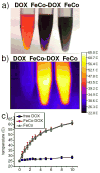
Figure 2. Photothermal heating of DOX and FeCo/GC Suspensions
(a) Image of solutions containing 450 μM DOX either as free DOX or FeCo/GC-DOX (FeCo-DOX). The FeCo/GC (FeCo) solution shown has the same FeCo/GC content as the FeCo/GC-DOX solution. (b) Thermal image after 10 minutes of 808 nm laser irradiation at 0.3 W/cm2. Both the FeCo/GC-DOX and FeCo/GC samples show significant photothermal heating, while the free DOX solution is not heated by the laser. (c) Temperature measurements from thermal images acquired during the 10 minute laser irradiation show a temperature increase of ~35°C for FeCo/GC containing samples. The free DOX sample showed almost no heating effect from laser irradiation.
Figure 3
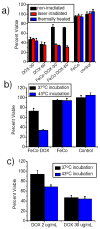
Figure 3. Cellular toxicity enhancement of DOX conjugates when combined with photothermal heating to 43°C
(a) Cell viability of MCF-7 cells after incubation with free DOX, FeCo/GC-DOX (FeCo-DOX), FeCo/GC (FeCo). DOX concentrations were either 30 or 60 μg DOX/mL. Samples were incubated for 20 minutes and combined with heating to 43°C through photothermal laser heating (red bars) or thermal heating in a water bath (blue bars). Non-heated samples are shown in black. FeCo/GC, free DOX or untreated control cells showed no viability dependence on the incubation conditions (heated vs. non-heated). FeCo/GC-DOX efficacy was significantly enhanced when incubations were performed at 43°C. The viability of control cells or FeCo/GC (NP) incubated cells were not affected by laser irradiation or thermal heating to 43°C for 20 minutes. (b) MCF-7 viability plots demonstrate the large increase in toxicity resulting from FeCo/GC-DOX (FeCo-DOX) at 43°C over 37°C, while cells incubated with FeCo/GC (FeCo) are not affected by thermal heating to 43°C for 20 minutes. (c) Cell toxicity of DOX at 37°C and 43°C reveals thermal enhancement of DOX toxicity at low doses of DOX (2 μg/mL). DOX efficacy of highly toxic doses (30 μg/mL) is not enhanced at 43°C due to the sufficiently high efficacy of high-dose DOX at 37°C.
Figure 4
Figure 4. Enhanced Cellular Uptake of FeCo/GC-DOX at 43°C
(a) Magnetic resonance image of pelletted, untreated MCF-7 cells or pelletted MCF-7 cells treated with FeCo/GC-DOX (FeCo-DOX) for 20 minutes at 43°C and 37°C. Brightening of samples in the T1-weighted images indicates FeCo/GC-DOX uptake. Cells incubated at 43°C with FeCo/GC-DOX appeared the brightest due to higher cellular uptake of FeCo/GC-DOX. (b) Region-of-interest (ROI) signal measurements of the cell pellets in part (a). MCF-7 cells treated with FeCo/GC-DOX had the highest signal intensity due to the higher uptake during the 20 minute incubation. (c) DOX fluorescence signal after being extracted from cells incubated with FeCo/GC-DOX (FeCo-DOX) for 20 minutes at 37°C or 43°C. (d) Flow cytometry measurements confirming higher DOX fluorescence in cells incubated with FeCo/GC-DOX at 43°C than at 37°C. Untreated MCF-7 cell signal is shown for comparison. The mean signal of cells treated with FeCo/GC-DOX for 20 minutes at 37°C versus 43°C is plotted in the inset. The ratio of the 43°C treatment versus the 37°C treatment is similar to the signal ratio in part (c).

References

    1. Purushotham S, Chang PEJ, Rumpel H, Kee IHC, Ng RTH, Chow PKH, Tan CK, Ramanujan RV. Thermoresponsive Core-Shell Magnetic Nanoparticles for Combined Modalities of Cancer Therapy. Nanotechnology. 2009;20:305101. - PubMed
    1. Maeng JH, Lee DH, Jung KH, Bae YH, Park IS, Jeong S, Jeon YS, Shim CK, Kim W, Kim J, et al. Multifunctional Doxorubicin Loaded Superparamagnetic Iron Oxide Nanoparticles for Chemotherapy and Magnetic Resonance Imaging in Liver Cancer. Biomaterials. 2010;31:4995–5006. - PubMed
    1. Park H, Yang J, Lee J, Haam S, Choi IH, Yoo KH. Multifunctional Nanoparticles for Combined Doxorubicin and Photothermal Treatments. ACS Nano. 2009;3:2919–2926. - PubMed
    1. Lee SM, Park H, Yoo KH. Synergistic Cancer Therapeutic Effects of Locally Delivered Drug and Heat Using Multifunctional Nanoparticles. Adv Mater. 2010;22:4049–53. - PubMed
    1. Torchilin VP. Targeted Pharmaceutical Nanocarriers for Cancer Therapy and Imaging. AAPS J. 2007;9:E128–E147. - PMC - PubMed

Publication types