

Observer variability in the interpretation of HER2/neu immunohistochemical expression with unaided and computer-aided digital microscopy
- PMID: 21284444
- PMCID: PMC7604903
- DOI: 10.5858/135.2.233
Observer variability in the interpretation of HER2/neu immunohistochemical expression with unaided and computer-aided digital microscopy
Abstract
Context: Observer variability in digital microscopy and the effect of computer-aided digital microscopy are underexamined areas in need of further research, considering the increasing use and future role of digital imaging in pathology. A reduction in observer variability using computer aids could enhance the statistical power of studies designed to determine the utility of new biomarkers and accelerate their incorporation in clinical practice.
Objectives: To quantify interobserver and intraobserver variability in immunohistochemical analysis of HER2/neu with digital microscopy and computer-aided digital microscopy, and to test the hypothesis that observer agreement in the quantitative assessment of HER2/neu immunohistochemical expression is increased with the use of computer-aided microscopy.
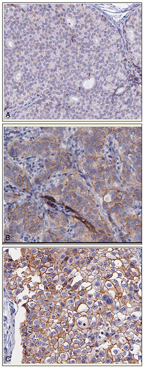
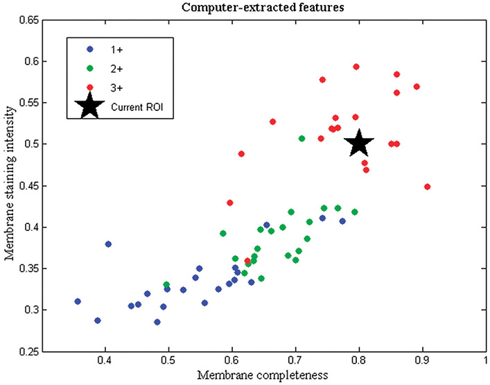
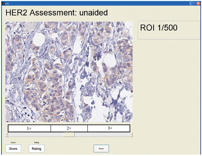
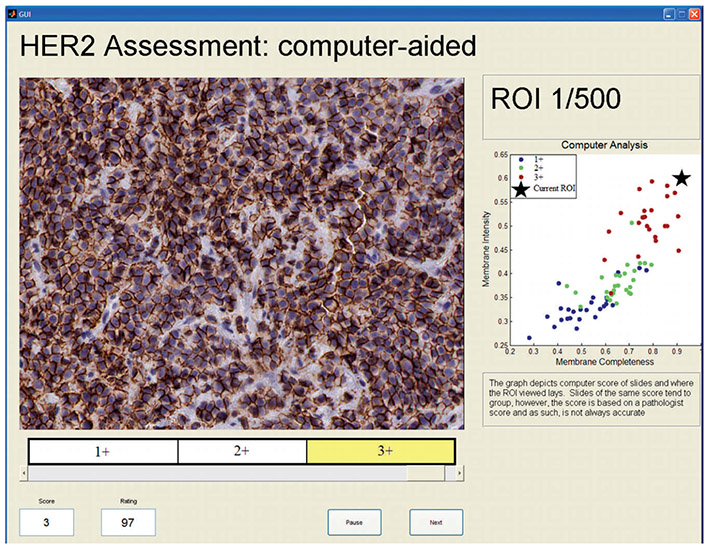
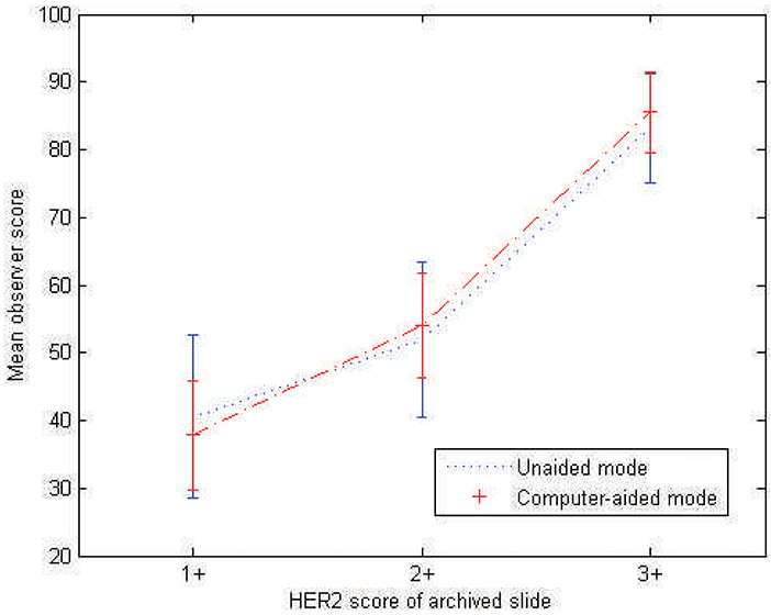
Design: A set of 335 digital microscopy images extracted from 64 breast cancer tissue slides stained with a HER2 antibody, were read by 14 observers in 2 reading modes: the unaided mode and the computer-aided mode. In the unaided mode, HER2 images were displayed on a calibrated color monitor with no other information, whereas in the computer-aided mode, observers were shown a HER2 image along with a corresponding feature plot showing computer-extracted values of membrane staining intensity and membrane completeness for the particular image under examination and, at the same time, mean feature values of the different HER2 categories. In both modes, observers were asked to provide a continuous score of HER2 expression.
Results: Agreement analysis performed on the output of the study showed significant improvement in both interobserver and intraobserver agreement when the computer-aided reading mode was used to evaluate preselected image fields.
Conclusion: The role of computer-aided digital microscopy in reducing observer variability in immunohistochemistry is promising.
Figures
References
-
- Taylor CR, Cote RJ. Immunomicroscopy, a Diagnostic Tool for the Surgical Pathologist. 3rd ed. Philadelphia, PA: Saunders Elsevier; 2006.
-
- Wolff AC, Hammond ME, Schwartz JN, et al. American Society of Clinical Oncology/College of American Pathologists guideline recommendations for human epidermal growth factor receptor 2 testing in breast cancer. J Clin Oncol. 2007;25(1):118–145. - PubMed
-
- Weinstein RS. Innovations in medical imaging and virtual microscopy. Hum Pathology. 2005;36(4):317–319. - PubMed
-
- Furness PN. The use of digital images in pathology. J Pathol. 1997;183(3):253–263. - PubMed
-
- Yagi Y, Gilbertson JR. Digital imaging in pathology: the case for standardization. J Telemed Telecare. 2005;11(3):109–116. - PubMed
Publication types
MeSH terms
Substances
Grants and funding
LinkOut - more resources
Full Text Sources
Medical
Research Materials
Miscellaneous