. 2011 Feb 1:10:27.
doi: 10.1186/1476-511X-10-27.

Purification and biochemical characterization of a secreted group IIA chicken intestinal phospholipase A₂

Affiliations

Purification and biochemical characterization of a secreted group IIA chicken intestinal phospholipase A₂

Aida Karray et al. Lipids Health Dis. .

Abstract

Background: Secretory phospholipase A2 group IIA (IIA PLA2) is a protein shown to be highly expressed in the intestine of mammals. However, no study was reported in birds.

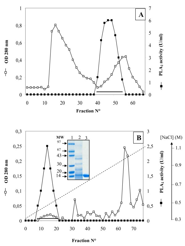
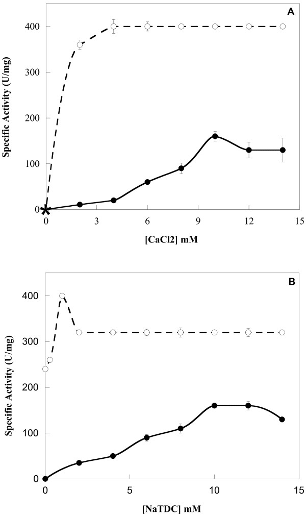
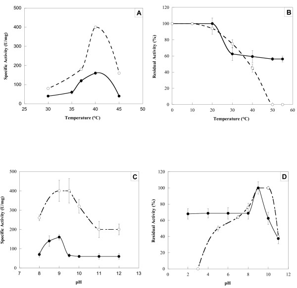
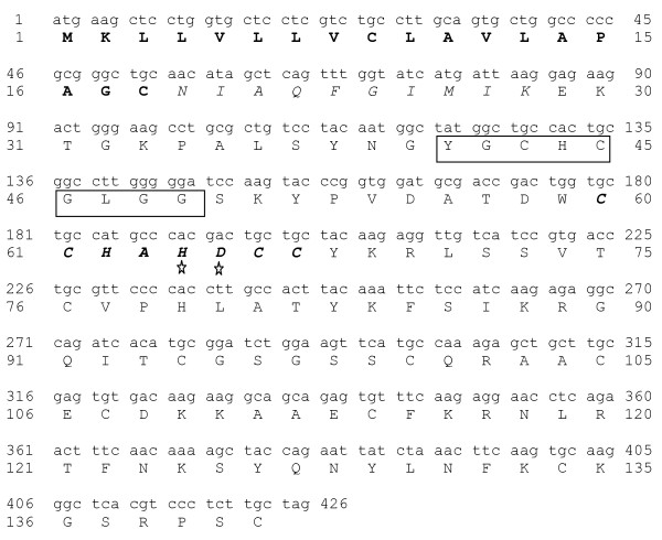
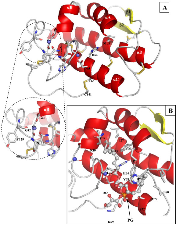
Results: Chicken intestinal group IIA phospholipase A₂ (ChPLA₂-IIA) was obtained after an acidic treatment (pH.3.0), precipitation by ammonium sulphate, followed by sequential column chromatographies on Sephadex G-50 and mono-S ion exchanger. The enzyme was found to be a monomeric protein with a molecular mass of around 14 kDa. The purified enzyme showed a substrate preference for phosphatidylethanolamine and phosphatidylglycerol, and didn't hydrolyse phosphatidylcholine. Under optimal assay conditions, in the presence of 10 mM NaTDC and 10 mM CaCl₂, a specific activity of 160 U.mg⁻¹ for purified ChPLA₂-IIA was measured using egg yolk as substrate. The fifteen NH2-terminal amino acid residues of ChPLA₂-IIA were sequenced and showed a close homology with known intestinal secreted phospholipases A₂. The gene encoding the mature ChPLA₂-IIA was cloned and sequenced. To further investigate structure-activity relationship, a 3D model of ChPLA₂-IIA was built using the human intestinal phospholipase A₂ structure as template.

Conclusion: ChPLA2-IIA was purified to homogeneity using only two chromatographic colomns. Sequence analysis of the cloned cDNA indicates that the enzyme is highly basic with a pI of 9.0 and has a high degree of homology with mammalian intestinal PLA₂-IIA.


Figures

Figure 1
Figure 1
Thin layer chromatography of PC and PE before or after incubation with ChPLA2-IIA or ChPLA2-IB. After solvent migration the silica plate was exposed to iodine vapor to reveal the various spots. Lane (1), PC. Lane (2), PC incubated with PLA2 from chicken pancreas (PLA2-IB) and showing a complete hydrolysis of the PC. Lane (3), PC incubated with PLA2 from chicken intestine (PLA2-IIA). Lane (4), PE. Lane (5), PE incubated with PLA2-IB and showing a complete hydrolysis of the PE. Lane (6), PE incubated with PLA2-IIA and showing a complete hydrolysis of the PE. Lane (7), free fatty acid C 18:1.
Figure 2
Figure 2
Purification of ChPLA2-IIA. (A) Gel filtration chromatography of intestinal ChPLA2-IIA on Sephadex G-50. The column (1.5 cm × 34 cm) equilibrated in 20 mM Tris-HCl buffer pH 8.0 containing 20 mM CaCl2 and 2 mM benzamidine. Elution was performed with the same buffer at a flow rate of 40 ml.h-1 and 3 ml samples were collected. ChPLA2-IIA activity was measured as described in Material and methods section using egg yolk emulsion as substrate. The pooled fractions containing the PLA2 activity were indicated by horizontal line. (B) Mono-S Sepharose chromatography. The column (5 cm × 2 cm) was equilibrated with 20 mM Tris HCl buffer pH 8.0 containing 20 mM CaCl2 and 2 mM benzamidine; and then washed with the same buffer containing 0.3 M NaCl. Linear salt gradient (0.3 to 1 M NaCl, dotted line) was applied to the column; gradient chamber 75 ml; 2 ml fraction; flow rate, 40 ml/h. The pooled fractions containing the PLA2 activity were indicated by horizontal line. SDS-PAGE (15%) analysis of pure ChPLA2-IIA was inserted in Figure 2B. Lane 1, molecular mass markers (MM); Lane 2, 15 μg of proteins obtained after Sephadex G-50 chromatography; Lane 3, 15 μg of purified ChPLA2-IIA, obtained after Mono-S chromatography.
Figure 3
Figure 3
Effect of Ca2+ and NaTDC on ChPLA2-IB and ChPLA2-IIA activities. (A) Effect of Ca2+ concentration on ChPLA2-IIA (black circle) and ChPLA2-IB (white circle) activities. Enzyme activity was measured at various concentrations of Ca2+ using egg yolk emulsion as substrate at pH 9.0 and at 40°C in the presence of 10 mM NaTDC. The star indicates the phospholipase activity measured in the absence of CaCl2 and in the presence of 10 mM EDTA or EGTA. (B) Effect of increasing concentration of bile salts (NaTDC) on ChPLA2-IIA (black circle) and ChPLA2-IB (white circle) activities. PLA2 activity was measured using egg yolk emulsion as substrate at pH 9.0 and at 40°C in the presence of 10 mM Ca2+.
Figure 4
Figure 4
Effect of temperature and pH on ChPLA2-IB and ChPLA2-IIA. Effects of temperature (A, B) and pH (C, D) on ChPLA2-IIA (black circle) and ChPLA2-IB (white circle) activities (A, C) and stability (B, D). PLA2 were tested for activity at various temperatures (A) and pH (C) as described in material and methods. To study the stability of PLA2, 1 mg.ml-1 of each enzyme was incubated during 30 min at various temperatures (B) and pH (D). Residual PLA2 activity was measured using egg yolk as substrate in the presence of 10 mM Ca2+ and 10 mM NaTDC. For temperature stability studies, enzymes were incubated in 10 mM Tris (pH 8.0) and 10 mM CaCl2. For pH stability studies, Tris buffer was replaced by the appropriate buffer for the pH range.
Figure 5
Figure 5
Nucleotide sequence of the cDNA of ChPLA2-IIA and the deduced amino acid sequence. Sequencing was performed in triplicate with three independent PCRs. The amino acid sequence obtained by N-terminal amino-acid sequence of the pure ChPLA2-IIA is shown in italic. M1-C18 in bold, signal peptide; Y41-G49 in square, Ca2+ loop; and H64-D65 with stars, active site.
Figure 6
Figure 6
3D modelling structure of ChPLA2-IIA. (A) Cartoon representation the 3D model of the mature ChPLA2-IIA. Secondary structure elements labels are indicated. α-helices and β-strands are colored in red and yellow, respectively. The catalytic network and the Ca2+-binding residues are indicated and shown as sticks. The disulfide-bridges are shown in yellow. (B) Cartoon representation of the ChPLA2-IIA structure showing amino acids (represented by sticks) interacting with the substrate (PG). The substrate is indicated as a stick representation. The Ca2+ ions are represented by a blue spheres. This figure was generated using the PYMOL software.

References

    1. Six DA, Dennis EA. The expanding superfamily of phospholipase A(2) enzymes: classification and characterization. Biochim Biophys Acta. 2000;1488(1-2):1–19. - PubMed
    1. Balsinde J, Balboa MA, Insel PA, Dennis EA. Regulation and inhibition of phospholipase A2. Annu Rev Pharmacol Toxicol. 1999;39:175–189. doi: 10.1146/annurev.pharmtox.39.1.175. - DOI - PubMed
    1. Valentin E, Lambeau G. Increasing molecular diversity of secreted phospholipases A(2) and their receptors and binding proteins. Biochim Biophys Acta. 2000;1488(1-2):59–70. - PubMed
    1. Nevalainen TJ, Haapanen TJ. Distribution of pancreatic (group I) and synovial-type (group II) phospholipases A2 in human tissues. Inflammation. 1993;17(4):453–464. doi: 10.1007/BF00916585. - DOI - PubMed
    1. Yoshikawa T, Naruse S, Kitagawa M, Ishiguro H, Nakae Y, Ono T, Hayakawa T. Effect of a new inhibitor of type II phospholipase A2 on experimental acute pancreatitis in rats. Pancreas. 1999;19(2):193–198. doi: 10.1097/00006676-199908000-00013. - DOI - PubMed

Publication types

MeSH terms