Review
. 2011 Jun 15;510(2):120-8.
doi: 10.1016/j.abb.2011.01.017. Epub 2011 Feb 1.

Myosin light chain kinase and the role of myosin light chain phosphorylation in skeletal muscle

Affiliations
Review

Myosin light chain kinase and the role of myosin light chain phosphorylation in skeletal muscle

James T Stull et al. Arch Biochem Biophys. .

Abstract

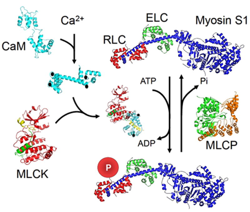
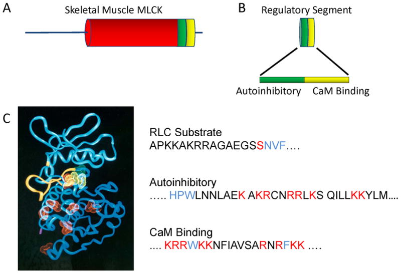
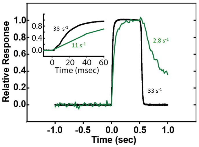
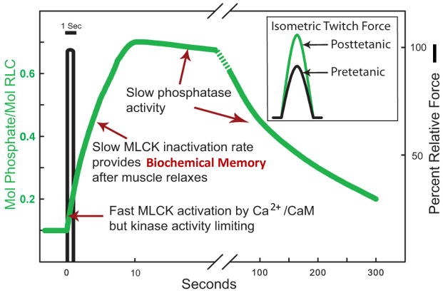
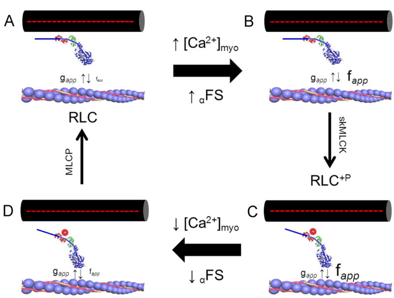
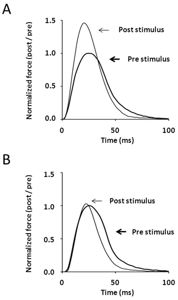
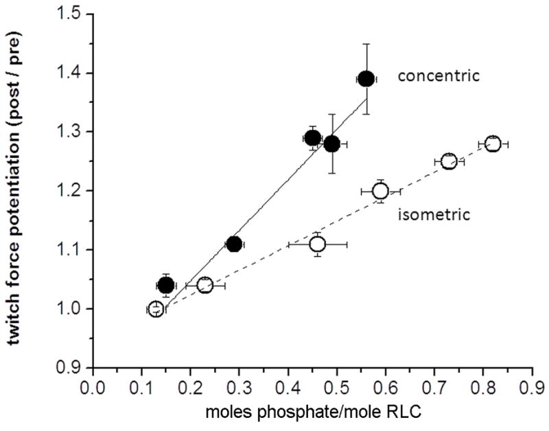
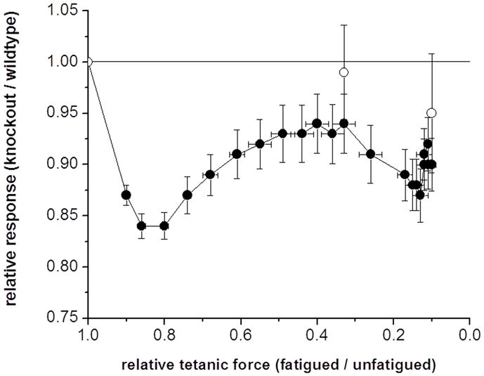
Skeletal muscle myosin light chain kinase (skMLCK) is a dedicated Ca(2+)/calmodulin-dependent serine-threonine protein kinase that phosphorylates the regulatory light chain (RLC) of sarcomeric myosin. It is expressed from the MYLK2 gene specifically in skeletal muscle fibers with most abundance in fast contracting muscles. Biochemically, activation occurs with Ca(2+) binding to calmodulin forming a (Ca(2+))(4)•calmodulin complex sufficient for activation with a diffusion limited, stoichiometric binding and displacement of a regulatory segment from skMLCK catalytic core. The N-terminal sequence of RLC then extends through the exposed catalytic cleft for Ser15 phosphorylation. Removal of Ca(2+) results in the slow dissociation of calmodulin and inactivation of skMLCK. Combined biochemical properties provide unique features for the physiological responsiveness of RLC phosphorylation, including (1) rapid activation of MLCK by Ca(2+)/calmodulin, (2) limiting kinase activity so phosphorylation is slower than contraction, (3) slow MLCK inactivation after relaxation and (4) much greater kinase activity relative to myosin light chain phosphatase (MLCP). SkMLCK phosphorylation of myosin RLC modulates mechanical aspects of vertebrate skeletal muscle function. In permeabilized skeletal muscle fibers, phosphorylation-mediated alterations in myosin structure increase the rate of force-generation by myosin cross bridges to increase Ca(2+)-sensitivity of the contractile apparatus. Stimulation-induced increases in RLC phosphorylation in intact muscle produces isometric and concentric force potentiation to enhance dynamic aspects of muscle work and power in unfatigued or fatigued muscle. Moreover, RLC phosphorylation-mediated enhancements may interact with neural strategies for human skeletal muscle activation to ameliorate either central or peripheral aspects of fatigue.


Figures

Fig. 1
Fig. 1
Scheme for myosin RLC phosphorylation in skeletal muscle based on structures of involved proteins. SkMLCK is inactive due to the regulatory segment containing autoinhibitory (green) and calmodulin-binding (yellow) sequences binding to the catalytic core. The phosphorylatable Ser in the N-terminus of RLC is thus prevented from binding in the catalytic cleft between the N-and C-domains of the catalytic core (red). Ca2+ (●) binds to four Ca2+ -binding sites in calmodulin (CaM), and the complex binds to skMLCK to displace the regulatory segment from the catalytic cleft. The subfragment 1 (S1) myosin head and neck domain comprises heavy chain (blue) with essential light chain (green, ECL) and RLC (red, RLC) bound to the α-helical neck region of the heavy chain. The disordered N-terminus of RLC (not shown) extends from RLC bound to the heavy chain for phosphorylation (P) by activated skMLCK. Myosin light chain phosphatase (MLCP) containing the catalytic subunit (green) bound to the regulatory subunit (orange) dephosphorylates phosphorylated RLC. The protein structures are discussed with references in the text.
Fig. 2
Fig. 2
Structural elements important for skMLCK. (A) SkMLCK has an N-terminal sequence followed by a catalytic core (red) and regulatory segment (green, yellow). (B) The regulatory segment has an autoinhibitory sequence immediately C-terminal of the catalytic core followed by the calmodulin-binding sequence. (C) Left panel: Acidic residues (red and yellow) predicted to be on the surface of the modeled catalytic core that may bind to the autoinhibitory sequence with yellow residues important to position RLC for phosphorylation. The N-terminus of RLC extends through the catalytic cleft binding to sites in the N-domain of the catalytic core (yellow ribbon). Right panel: The primary substrate determinants in the N-terminus of skeletal muscle RLC are shown in blue relative to the phosphorylatable Ser (red). The autoinhibitory sequence of skMLCK extends from the catalytic core (blue residues) with basic residues (red) binding to acidic residues on the surface of the catalytic core. Ca2+/calmodulin collapses around the calmodulin-binding sequence containing two hydrophobic residues (blue) with additional ionic interactions with basic residues (red).
Fig. 3
Fig. 3
SkMLCK is rapidly activated (green) by Ca2+/calmodulin in skeletal muscle stimulated to develop force (black) but inactivated more slowly relative to relaxation [43]. The slower inactivation rate provides a biochemical memory effect for RLC phosphorylation.
Fig. 4
Fig. 4
Biochemical determinants for physiological phosphorylation of RLC in fast skeletal muscle. A brief tetanic contraction results in RLC phosphorylation related to the amount of fully activated skMLCK in muscle fibers, with slow inactivation due to the slow dissociation of Ca2+/calmodulin from the kinase after relaxation. RLC phosphorylation persists for some time after the skMLCK is inactivated due to low phosphatase activity. The insert shows isometric twitch force responses without (black) and with (green) RLC phosphorylation.
Fig. 5
Fig. 5
RLC phosphorylation affects myosin cross bridge structure and function during Ca2+ activation of skeletal muscle contraction. In this model, the regulatory influence of Ca2+ on force development is exerted via changes in the rate constant describing the transition of cycling cross bridges from non-force to force-generating states (i.e. fapp). An increase in myoplasmic [Ca2+] ([Ca2+]myo) and concomitant thin filament activation increases force by regulating the fraction of cycling crossbridges able to accumulate in force-generating states (αFS). The addition of a negatively charged phosphate to Ser15 in RLC (red circle) displaces the myosin motor domain laterally from the thick filament towards thin filament binding sites, which affects Ca2+ control of cross bridge kinetics. Clockwise from top left: (A) unphosphorylated cross bridge at rest; when myoplasmic [Ca2+] is low the transition of strong to weak binding states (i.e. gapp) dominates and force is low; (B) unphosphorylated cross bridge during contraction in response to elevated myoplasmic [Ca2+] showing Ca2+ regulated increase in fapp; gapp is not influenced; (C) phosphorylated crossbridge during contraction; the modulation of fapp further increases the fraction of cross bridges able to attain force generating states to increase the Ca2+ sensitivity of force development relative to unphosphorylated state; (D) phosphorylated crossbridge during relaxation; the return of myoplasmic [Ca2+] to low levels may be accompanied by a small residual increase in fapp to slow the rate of relaxation from contraction. Small upward and downward vertical arrows denote the terms fapp and gapp, respectively. MLCP, myosin light chain phosphatase. Based on references [67, 78]
Fig. 6
Fig. 6
Comparison of isometric twitch responses in EDL muscles from wild-type (A) and skMLCK knockout (B) mice obtained 1 minute before and 15 sec after a potentiating stimulus consisting of four brief trains of 150-Hz stimulation (within 10 sec) that elevated myosin RLC phosphorylation from 0.15 to 0.55 mol phosphate per mol RLC. Note the large (~ 50% post/pre) increase in twitch amplitude of the wild-type muscle relative to the skMLCK knockout muscle. Experiments performed at 25° C with muscles set to 0.90 of optimal length (Lo).
Fig. 7
Fig. 7
Relation of concentric and isometric twitch force potentiation versus RLC phosphorylation obtained following stimulation at different frequencies (2.5 to 100 Hz). Each data set fitted with linear regression. Concentric (closed circles) data are unpublished; isometric (open circles) data re-plotted from Vandenboom et al.[85]. Note that despite similar y-intercepts, the concentric force – phosphorylation relationship is steeper than is the isometric force - phosphorylation relationship. Data are means ± SEM (n=4–8 muscles each time point).
Fig. 8
Fig. 8
Plot of relative change in low frequency force (Pt) (closed symbols) and unloaded shortening velocity (Vo) (open symbols) measured via the slack test method versus tetanic force decline in muscles from wild-type and skMLCK knockout mice. Muscles were stimulated for 5 minutes during which time Po was reduced to ~ 10% of starting levels in each muscle type. Vo was determined before, during (after 1 minute of stimulation) and immediately after the fatigue run. All data obtained by dividing responses from muscles from skMLCK knockout to wild-type mice and plotted against the corresponding change in tetanic force (the same for muscles from skMLCK knockout and wild-type mice). The Pt of skMLCK KO muscles was reduced more than was the Pt of WT muscles (= 1.00) for all fatigue levels. On the other hand, the Vo of skMLCK KO muscles was similar to the Vo of WT muscles (=1.00) for all levels of fatigue. Data is mean ± SEM (n=12 for each genotype). Muscles were set to 0.90 Lo and subjected to 5 minutes of repetitive high frequency stimulation (150 Hz) (redrawn with permission [98].

References

    1. Gordon AM, Homsher E, Regnier M. Physiol Rev. 2000;80:853–924. - PubMed
    1. Rayment I. J Biol Chem. 1996;271:15850–15853. - PubMed
    1. Lowey S, Trybus KM. Journal of Biological Chemistry. 2010;285:16403–16407. - PMC - PubMed
    1. Kamm KE, Stull JT. J Biol Chem. 2001;276:4527–4530. - PubMed
    1. Manning G, Whyte DB, Martinez R, Hunter T, Sudarsanam S. Science. 2002;298:1912–1934. - PubMed

Publication types

MeSH terms

Substances

LinkOut - more resources