Drosophila UNC-45 accumulates in embryonic blastoderm and in muscles, and is essential for muscle myosin stability
- PMID: 21285246
- PMCID: PMC3039016
- DOI: 10.1242/jcs.078964
Drosophila UNC-45 accumulates in embryonic blastoderm and in muscles, and is essential for muscle myosin stability
Abstract
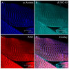
UNC-45 is a chaperone that facilitates folding of myosin motor domains. We have used Drosophila melanogaster to investigate the role of UNC-45 in muscle development and function. Drosophila UNC-45 (dUNC-45) is expressed at all developmental stages. It colocalizes with non-muscle myosin in embryonic blastoderm of 2-hour-old embryos. At 14 hours, it accumulates most strongly in embryonic striated muscles, similarly to muscle myosin. dUNC-45 localizes to the Z-discs of sarcomeres in third instar larval body-wall muscles. We produced a dunc-45 mutant in which zygotic expression is disrupted. This results in nearly undetectable dUNC-45 levels in maturing embryos as well as late embryonic lethality. Muscle myosin accumulation is robust in dunc-45 mutant embryos at 14 hours. However, myosin is dramatically decreased in the body-wall muscles of 22-hour-old mutant embryos. Furthermore, electron microscopy showed only a few thick filaments and irregular thick-thin filament lattice spacing. The lethality, defective protein accumulation, and ultrastructural abnormalities are rescued with a wild-type dunc-45 transgene, indicating that the mutant phenotypes arise from the dUNC-45 deficiency. Overall, our data indicate that dUNC-45 is important for myosin accumulation and muscle function. Furthermore, our results suggest that dUNC-45 acts post-translationally for proper myosin folding and maturation.
Figures


Similar articles
-
The UNC-45 chaperone is critical for establishing myosin-based myofibrillar organization and cardiac contractility in the Drosophila heart model.PLoS One. 2011;6(7):e22579. doi: 10.1371/journal.pone.0022579. Epub 2011 Jul 25. PLoS One. 2011. PMID: 21799905 Free PMC article.
-
Drosophila UNC-45 prevents heat-induced aggregation of skeletal muscle myosin and facilitates refolding of citrate synthase.Biochem Biophys Res Commun. 2010 May 28;396(2):317-22. doi: 10.1016/j.bbrc.2010.04.090. Epub 2010 Apr 18. Biochem Biophys Res Commun. 2010. PMID: 20403336 Free PMC article.
-
Knockdown and overexpression of Unc-45b result in defective myofibril organization in skeletal muscles of zebrafish embryos.BMC Cell Biol. 2010 Sep 17;11:70. doi: 10.1186/1471-2121-11-70. BMC Cell Biol. 2010. PMID: 20849610 Free PMC article.
-
The UNC-45 myosin chaperone: from worms to flies to vertebrates.Int Rev Cell Mol Biol. 2014;313:103-44. doi: 10.1016/B978-0-12-800177-6.00004-9. Int Rev Cell Mol Biol. 2014. PMID: 25376491 Free PMC article. Review.
-
Determining structure/function relationships for sarcomeric myosin heavy chain by genetic and transgenic manipulation of Drosophila.Microsc Res Tech. 2000 Sep 15;50(6):430-42. doi: 10.1002/1097-0029(20000915)50:6<430::AID-JEMT2>3.0.CO;2-E. Microsc Res Tech. 2000. PMID: 10998634 Review.
Cited by
-
Hsf1 is essential for proteotoxic stress response in smyd1b-deficient embryos and fish survival under heat shock.FASEB J. 2025 Jan 15;39(1):e70283. doi: 10.1096/fj.202401875R. FASEB J. 2025. PMID: 39760245
-
UNC-45A: A potential therapeutic target for malignant tumors.Heliyon. 2024 May 14;10(10):e31276. doi: 10.1016/j.heliyon.2024.e31276. eCollection 2024 May 30. Heliyon. 2024. PMID: 38803956 Free PMC article. Review.
-
Disruption of Drosophila larval muscle structure and function by UNC45 knockdown.BMC Mol Cell Biol. 2021 Jul 13;22(1):38. doi: 10.1186/s12860-021-00373-7. BMC Mol Cell Biol. 2021. PMID: 34256704 Free PMC article.
-
UNC-45A breaks the microtubule lattice independently of its effects on non-muscle myosin II.J Cell Sci. 2021 Jan 8;134(1):jcs248815. doi: 10.1242/jcs.248815. J Cell Sci. 2021. PMID: 33262310 Free PMC article.
-
UFD-2 is an adaptor-assisted E3 ligase targeting unfolded proteins.Nat Commun. 2018 Feb 2;9(1):484. doi: 10.1038/s41467-018-02924-7. Nat Commun. 2018. PMID: 29396393 Free PMC article.
References
-
- Adams M. D., Celniker S. E., Holt R. A., Evans C. A., Gocayne J. D., Amanatides P. G., Scherer S. E., Li P. W., Hoskins R. A., Galle R. F. (2000). The genome sequence of Drosophila melanogaster. Science 287, 2185-2195 - PubMed
-
- Atkinson S. J., Stewart M. (1991). Expression in Escherichia coli of fragments of the coiled-coil rod domain of rabbit myosin: influence of different regions of the molecule on aggregation and paracrystal formation. J. Cell Sci. 99, 823-836 - PubMed
Publication types
MeSH terms
Substances
Grants and funding
LinkOut - more resources
Full Text Sources
Molecular Biology Databases

