

MG53 participates in ischaemic postconditioning through the RISK signalling pathway
- PMID: 21285295
- PMCID: PMC3112015
- DOI: 10.1093/cvr/cvr029
MG53 participates in ischaemic postconditioning through the RISK signalling pathway
Abstract
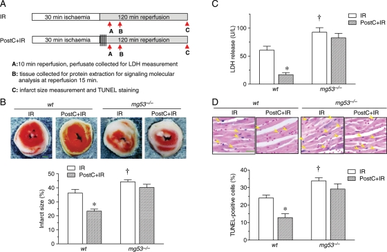
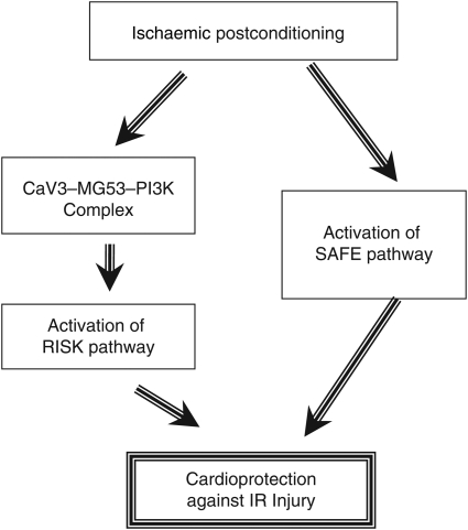
Aims: Recent studies show that ischaemic postconditioning (PostC), similar to the well-established ischaemic preconditioning (IPC), confers cardioprotection against ischaemia/reperfusion (IR) injury, and both IPC and PostC can activate the reperfusion injury salvage kinase (RISK) pathway and the survivor activating factor enhancement (SAFE) pathway. PostC is clinically more attractive because of its therapeutic application at the predictable onset of reperfusion. Our previous studies have demonstrated that MG53 is a primary component of the IPC machinery. Here, we investigated the potential role of MG53 in PostC-mediated myocardial protection and explored the underlying mechanism.
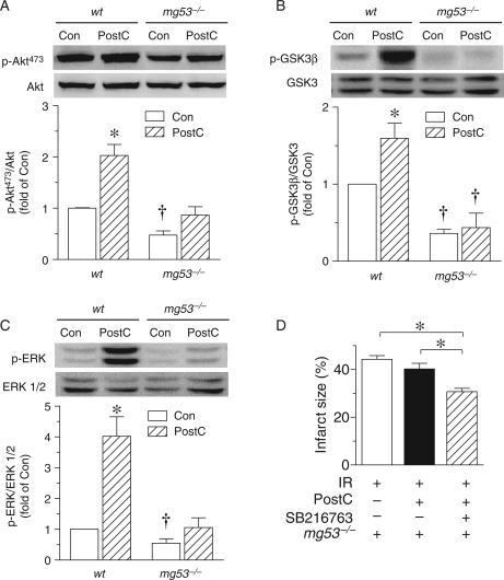
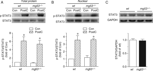
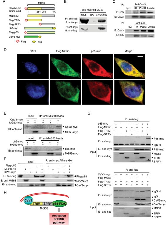
Methods and results: Using Langendorff perfusion, we investigated IR injury in wild-type (wt) and MG53-deficient (mg53(-/-)) mouse hearts with or without PostC. IR-induced myocardial damage was markedly exacerbated in mg53(-/-) hearts compared with wt controls. PostC protected wt hearts against IR-induced myocardial infarction, myocyte necrosis, and apoptosis, but failed to protect mg53(-/-) hearts. The loss of PostC protection in mg53(-/-) hearts was attributed to selectively impaired PostC-activated RISK signalling. Mechanistically, MG53 is required for the interaction between caveolin 3 (CaV3) and the p85 subunit of phosphoinositide 3-kinase (p85-PI3K) and PostC-mediated activation of the RISK pathway. Importantly, a structure-function study revealed that the MG53 tripartite motif (TRIM) domain (aa1-284) physically interacted with CaV3 but not p85-PI3K, whereas the MG53 SPRY domain (aa285-477) interacted with p85-PI3K but not CaV3, indicating that MG53 binds to CaV3 and p85 at its N- and C-terminus, respectively.
Conclusions: We conclude that MG53 participates in PostC-mediated cardioprotection largely through tethering CaV3 and PI3K and subsequent activation of the RISK pathway.
Figures
References
-
- Murry CE, Jennings RB, Reimer KA. Preconditioning with ischemia: a delay of lethal cell injury in ischemic myocardium. Circulation. 1986;74:1124–1136. - PubMed
-
- Zhao ZQ, Corvera JS, Halkos ME, Kerendi F, Wang NP, Guyton RA, et al. Inhibition of myocardial injury by ischemic postconditioning during reperfusion: comparison with ischemic preconditioning. Am J Physiol Heart Circ Physiol. 2003;285:H579–H588. - PubMed
-
- Lacerda L, Somers S, Opie LH, Lecour S. Ischaemic postconditioning protects against reperfusion injury via the SAFE pathway. Cardiovasc Res. 2009;84:201–208. - PubMed
-
- Tsang A, Hausenloy DJ, Mocanu MM, Yellon DM. Postconditioning: a form of “modified reperfusion” protects the myocardium by activating the phosphatidylinositol 3-kinase-Akt pathway. Circ Res. 2004;95:230–232. - PubMed
-
- Argaud L, Gateau-Roesch O, Raisky O, Loufouat J, Robert D, Ovize M. Postconditioning inhibits mitochondrial permeability transition. Circulation. 2005;111:194–197. - PubMed
Publication types
MeSH terms
Substances
LinkOut - more resources
Full Text Sources
Other Literature Sources
Medical
Molecular Biology Databases
