. 2011 Feb;23(2):534-49.
doi: 10.1105/tpc.110.081059. Epub 2011 Feb 1.

The R2R3 MYB transcription factor DUO1 activates a male germline-specific regulon essential for sperm cell differentiation in Arabidopsis

Affiliations

The R2R3 MYB transcription factor DUO1 activates a male germline-specific regulon essential for sperm cell differentiation in Arabidopsis

Michael Borg et al. Plant Cell. 2011 Feb.

Abstract

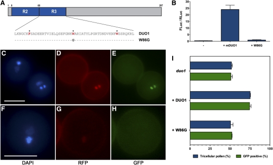
The male germline in flowering plants arises through asymmetric division of a haploid microspore. The resulting germ cell undergoes mitotic division and specialization to produce the two sperm cells required for double fertilization. The male germline-specific R2R3 MYB transcription factor DUO1 POLLEN1 (DUO1) plays an essential role in sperm cell specification by activating a germline-specific differentiation program. Here, we show that ectopic expression of DUO1 upregulates a significant number (~63) of germline-specific or enriched genes, including those required for fertilization. We validated 14 previously unknown DUO1 target genes by demonstrating DUO1-dependent promoter activity in the male germline. DUO1 is shown to directly regulate its target promoters through binding to canonical MYB sites, suggesting that the DUO1 target genes validated thus far are likely to be direct targets. This work advances knowledge of the DUO1 regulon that encompasses genes with a range of cellular functions, including transcription, protein fate, signaling, and transport. Thus, the DUO1 regulon has a major role in shaping the germline transcriptome and functions to commit progenitor germ cells to sperm cell differentiation.


Figures

Figure 1.
Figure 1.
MYB Binding Sites Are Overrepresented in DUO1 Target Gene Promoters. (A) DUO1 candidate targets divided into three groups based on their response time to ectopic DUO1 expression. Group A genes (black) have a 3-fold increase after 6 h of DUO1 induction, Group B (blue) after 12 h, and Group C (red) only after 24 h of induction. (B) Bioinformatic analysis of the promoters of candidate DAT genes identified three overrepresented MYB binding motifs. The consensus sequence of the motifs is represented as a PSSM logo. (C) A feature map encompassing positional information of the overrepresented MYB binding motifs in the 17 validated DAT gene promoters. The colored boxes indicate the position of each MYB binding site with each color corresponding to the PSSM logos in (B).
Figure 2.
Figure 2.
A Selection of Induced Target Genes Exhibit DUO1-Dependent Expression in the Male Germline. (A) Promoter activity of male germline-specific DAT genes. GFP expression was scored in single insert hemizygous lines in the duo1-1/+ background. Wild-type (WT) pollen grains (black) were distinguished from duo1 pollen grains (white) by germ cell number. Each bar represents the mean of at least three independent lines and error bars represent the se. All 12 promoters show significantly reduced activity in duo1 germ cells compared with wild-type sperm cells (two-tailed χ2 test: P < 0.0001). (B) to (D) Examples of male germline-specific DAT gene promoter activity in wild-type and duo1 pollen using CLSM. Each panel shows a representative pollen grain under DAPI fluorescence (left) and GFP fluorescence (right). Sperm cell nuclei in wild-type pollen grains show a GFP signal (B), while duo1 germ cell nuclei show a residual level (C) or no detectable GFP signal (D). Arrowheads indicate a residual level of GFP signal in duo1 germ cells. Bars = 10 μm. (E) Promoter activity of DAT genes expressed in the vegetative and sperm cells. GFP expression was scored in single insert hemizygous lines in the duo1-1/+ background for wild-type sperm cells (black), wild-type vegetative nuclei (dark gray), duo1 germ cells (white), and duo1 vegetative nuclei (light gray). Each bar chart represents the mean of at least three independent lines, and error bars show the se. The two promoters show significantly reduced activity in duo1 germ cells compared with wild-type sperm cells (two-tailed χ2 test: P < 0.0001), but there is no significant difference between activity in the vegetative cells. (F) to (H) Examples of non-male germline-specific DAT gene promoter activity in wild-type and duo1 pollen grains using CLSM. Each panel shows a representative pollen grain under DAPI fluorescence (left) and GFP fluorescence (right). Both sperm cell and vegetative cell nuclei in wild-type pollen grains show a GFP signal (F), while duo1 germ cell nuclei either show a residual level (G) or no detectable GFP signal (H), and the GFP signal of vegetative cell nuclei is unaffected in duo1 pollen grains ([G] and [H]). Arrowheads indicate a residual level of GFP signal in duo1 germ cells. Bars = 10 μm. (I) DUO1-dependent transactivation of validated DAT promoters in tobacco leaves. The relative luciferase activity (FLuc/RLuc) of each target promoter (ProDAT:LUC) alone (−; light gray) and upon coinfiltration with Pro35S:mDUO1 (+; dark gray) is shown with the fold change indicated above the (+) column. Each bar represents the mean of at least four independent infiltrations, and error bars show the se. A split y axis is presented in order to illustrate lower level activity. [See online article for color version of this figure.]
Figure 3.
Figure 3.
Promoter Activity of DUO1 Target Genes during Male Gametogenesis Expression of ProTIP5;1:H2B-GFP ([A] to [D]), ProIMPa-8:H2B-GFP ([E] to [H]), ProDAA1:H2B-GFP ([I] to [L]), and ProPCR11:H2B-GFP ([M] to [P]) during wild-type pollen development as observed with fluorescence microscopy. Representative pollen grains show GFP fluorescence (top, green) and DAPI fluorescence (bottom, blue) at microspore (MSP), early bicellular (EBC), late bicellular (LBC), and mature pollen (MPG) stages. Bars = 10 μm.
Figure 4.
Figure 4.
MYB Binding Sites Are Critical for DUO1-Dependent Transactivation of the MGH3 Promoter. (A) Relative luciferase activity of 5′ deletions in the MGH3 promoter. Left shows the promoter sequences highlighting MYB sites (black boxes) and the canonical TATA box (white box). Right shows the mean relative luciferase activity (FLuc/RLuc) for at least four independent infiltrations, and error bars show the se. (B) The significance of MYB sites for DUO1-dependent transactivation of the MGH3 promoter. Left shows the promoter fragments with targeted mutations and/or deletions in context of d3 of the MGH3 promoter with MYB sites named A to D. Right shows the mean FLuc/RLuc for at least four independent infiltrations, and error bars show the se. (C) Example of ProMGH3d3:H2B-GFP activity in mature pollen. Bar = 15 μm. (D) Example of ProMGH3d3_mAB:H2B-GFP activity in mature pollen. A clear decrease in GFP signal was observed in several independent lines. Error bars represent the se. Bar = 15 μm. (E) The decrease in GFP signal observed in ProMGH3d3_mAB:H2B-GFP lines. GFP fluorescence represents the mean total pixel intensity corrected for background in (n) sperm cells analyzed. Asterisks indicate statistically significant differences determined using a two-tailed Mann-Whitney U test (P < 0.0001, U = 2768).
Figure 5.
Figure 5.
DUO1 Binds in Vitro to a MYB Binding Site in the MGH3 Promoter. (A) Sequences of the oligos used in the EMSA experiments. MBS is the region flanking MYB site A in the MGH3 promoter (bold and underlined), and mMBS is a mutagenized version in which the MYB site has been ablated (underlined). (B) EMSA experiments using recombinant DUO1 MYB domain with MBS or mMBS oligos. – and + indicate the absence or presence of the DUO1 MYB domain, respectively, and ×125, ×250, and ×250 indicate competing unlabeled oligos. Addition of DUO1 MYB protein (lane 2 to 5) causes a clear shift (white triangle) accompanied by a reduction in free probes (black triangle), with free probes increasing in the presence of more competing MBS oligos. In lanes 7 to 10, no shift was observed in EMSA experiments using mMBS oligos. Results were confirmed in independent EMSA experiments.
Figure 6.
Figure 6.
DNA Binding Integrity of the R3R3 MYB Domain Is Critical for DUO1 Function. (A) Schematic diagram of the DUO1 protein with the R2 and R3 MYB repeats shown in blue. Below is the amino acid sequence of the R3 MYB repeat, with the red and asterisked amino acids representing the hydrophobic scaffold. Below the native sequence (DUO1) is the variant sequence (W86G) showing the mutation that compromises DNA binding capacity. (B) Transactivation of the MGH3 promoter by mDUO1 and mW86G. Bars show the relative luciferase activity (Fluc/Rluc) of ProMGH3:LUC in tobacco leaves with and without coinfiltration of Pro35S:mDUO1 and Pro35S:mW86G in at least four independent infiltrations, with error bars showing the se. (C) to (H) Mature pollen from transgenic lines expressing ProDUO1:DUO1-mRFP and ProDUO1:W86G-mRFP in duo1/+ plants homozygous for a ProMGH3:H2B-GFP marker. Each panel from left to right shows the same pollen grains with DAPI, mRFP, and GFP fluorescence as illustrated below the images. (C) to (E) Top left is an unrescued duo1 pollen grain with a bicellular phenotype (C) and no expression of the MGH3 marker (E). Bottom right is a fully rescued pollen grain, with expression of ProDUO1:DUO1-mRFP (D) restoring tricellularity (C) and MGH3 marker expression (E). (F) to (H) duo1 pollen grains expressing ProDUO1:W86G-mRFP (G) remain bicellular (F) and do not express the MGH3 marker (H). (I) Ability of ProDUO1:DUO1-mRFP and ProDUO1:W86G-mRFP to rescue germ cell division (blue column) and MGH3 transactivation (green column) in duo1 pollen. In duo1/+ plants (top columns), ~50% of the pollen is wild type and has two GFP-positive sperm cells. Expression of DUO1-mRFP (middle columns) rescues both germ cell division and activates MGH3 expression in the 50% of duo1 germ cells (25% total pollen) that contain the transgene. Expression of W86G-mRFP (bottom columns) is unable to rescue germ cell division or transactivate the MGH3 marker line. Bars show the mean of at least four hemizygous single locus lines, and error bars represent the se. [See online article for color version of this figure.]

References

    1. Baek K.H. (1999). The first oncogene in Drosophila melanogaster. Mutat. Res. 436: 131–136 - PubMed
    1. Bell S.D., Botting C.H., Wardleworth B.N., Jackson S.P., White M.F. (2002). The interaction of Alba, a conserved archaeal chromatin protein, with Sir2 and its regulation by acetylation. Science 296: 148–151 - PubMed
    1. Berger F., Hamamura Y., Ingouff M., Higashiyama T. (2008). Double fertilization - caught in the act. Trends Plant Sci. 13: 437–443 - PubMed
    1. Bilder D. (2001). Cell polarity: Squaring the circle. Curr. Biol. 11: R132–R135 - PubMed
    1. Borges F., Gomes G., Gardner R., Moreno N., McCormick S., Feijó J.A., Becker J.D. (2008). Comparative transcriptomics of Arabidopsis sperm cells. Plant Physiol. 148: 1168–1181 - PMC - PubMed

Publication types

MeSH terms