. 2011 Apr 1;286(13):11265-74.
doi: 10.1074/jbc.M110.182097. Epub 2011 Feb 1.

Protein kinase D negatively regulates hepatitis C virus secretion through phosphorylation of oxysterol-binding protein and ceramide transfer protein

Affiliations

Protein kinase D negatively regulates hepatitis C virus secretion through phosphorylation of oxysterol-binding protein and ceramide transfer protein

Yutaka Amako et al. J Biol Chem. .

Abstract

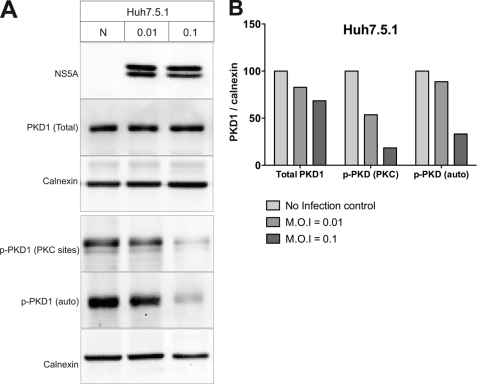
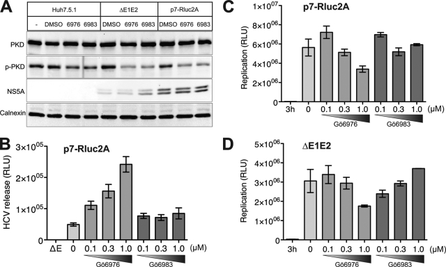
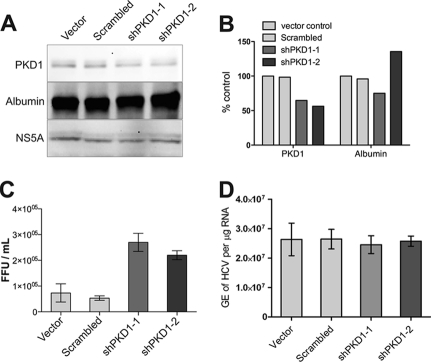
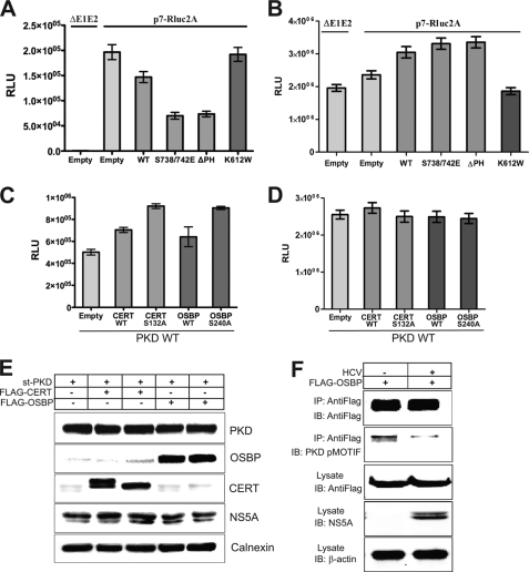
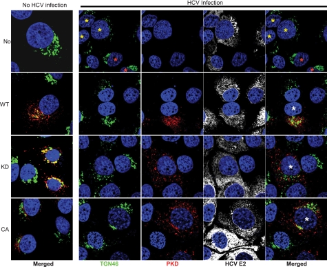
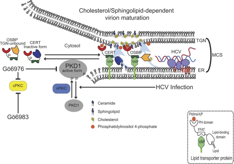
Hepatitis C virus (HCV) RNA replicates its genome on specialized endoplasmic reticulum modified membranes termed membranous web and utilizes lipid droplets for initiating the viral nucleocapsid assembly. HCV maturation and/or the egress pathway requires host sphingolipid synthesis, which occur in the Golgi. Ceramide transfer protein (CERT) and oxysterol-binding protein (OSBP) play a crucial role in sphingolipid biosynthesis. Protein kinase D (PKD), a serine/threonine kinase, is recruited to the trans-Golgi network where it influences vesicular trafficking to the plasma membrane by regulation of several important mediators via phosphorylation. PKD attenuates the function of both CERT and OSBP by phosphorylation at their respective Ser(132) and Ser(240) residues (phosphorylation inhibition). Here, we investigated the functional role of PKD in HCV secretion. Our studies show that HCV gene expression down-regulated PKD activation. PKD depletion by shRNA or inhibition by pharmacological inhibitor Gö6976 enhanced HCV secretion. Overexpression of a constitutively active form of PKD suppressed HCV secretion. The suppression by PKD was subverted by the ectopic expression of nonphosphorylatable serine mutant CERT S132A or OSBP S240A. These observations imply that PKD negatively regulates HCV secretion/release by attenuating OSBP and CERT functions by phosphorylation inhibition. This study identifies the key role of the Golgi components in the HCV maturation process.


Figures

FIGURE 1.
FIGURE 1.
HCV infection down-regulates PKD1 activation in Huh7.5.1 cells. A, analysis of PKD expression in HCV-infected cells. Huh7.5.1 cells were infected with culture-derived HCV particles by the indicated m.o.i. Six days after infection, whole cell lysates were analyzed by Western blot assay. Phosphorylated serine residues at 738 and 742 (PKC sites) or 910 (autophosphorylation site) were probed using phosphoserine substrate-specific antibody. Both phosphorylations represent the enzymatically active form of PKD1 (p-PKD1). B, quantitation of PKD1 expression and activation. Detected protein band intensities (shown in A) were calculated by 1D Image Analysis Software (Eastman Kodak Co.). PKD protein band intensity was normalized to calnexin. The values are represented as percentages relative to uninfected control (N).
FIGURE 2.
FIGURE 2.
PKD inhibitor Gö6976 promotes HCV secretion in dose-dependent manner. A, effect of PKD/PKC (Gö6976) and PKC (Gö6983) inhibitors on protein expression as shown by Western blot assays. Huh7.5.1 cells were mock-transfected or transfected with in vitro transcribed p7-Rluc2A RNA as indicated. Transfected cells were treated with or without 1 μm Gö6976 or Gö6983 2 days after transfection and incubated for 3 additional days. Electroblotted membranes were probed with the following antibodies: anti-PKD, anti-phospho-PKD1 (p-PKD; PKC sites), anti-NS5A, and anti-calnexin. B, effect of PKC (Gö6983) and PKC/PKD (Gö6976) inhibitors on HCV virion release. Cultured supernatants were used to infect naïve Huh7.5.1 cells, and cellular lysates were evaluated for Renilla luciferase activity. This protocol detects the HCV virion release. The E1E2 mutant (ΔE), which contains an in-frame deletion within the E1 and E2 coding sequence and hence is incapable of producing infectious virus particles, was used as a negative control in the HCV virion release assay. C, effect of PKC (Gö6983) and PKC/PKD (Gö6976) inhibitors on HCV replication. Intracellular luciferase activities were measured for samples as described in B. This analysis represents intracellular viral RNA replication. D, effect of PKC (Gö6983) and PKC/PKD (Gö6976) inhibitors on HCV replication. p7-Rluc2A containing the E1E2 deletion (ΔE1E2) was used to monitor RNA replication in the presence of inhibitors. This mutant is defective in HCV particle formation. All experiments are carried out in triplicate and the data shown are mean ± S.E. RLU, relative luciferase units.
FIGURE 3.
FIGURE 3.
PKD1 depletion promotes HCV secretion. Huh7.5.1 cells were transfected with plasmid vectors encoding shRNA as indicated. Four days after transfection, cells were electroporated with in vitro transcribed HCV JC1 RNA and incubated for 3 days. Whole cell lysates were subjected to Western blot assays (A). Panels represent expression of total PKD1, albumin, and HCV NS5A, respectively. B, the level of PKD1 depletion was quantified by 1D Image Analysis Software (Kodak). C, HCV secretion/release was measured by the focus-forming unit (FFU) assay as described under “Experimental Procedures.” D, HCV replication determined by quantitative RT-PCR analysis of HCV genome equivalents (GE)/μg of total intracellular RNA. The data shown are mean ± S.E. of three independent experiments.
FIGURE 4.
FIGURE 4.
PKD regulates HCV secretion via phosphorylation of CERT and OSBP. A, effect of PKD overexpression on HCV secretion. Huh7.5.1 cells were infected with lentiviral vectors encoding PKD1 (WT), dominant active (S738/742E), PH domain deletion (ΔPH) mutant, and kinase-inactive mutant (K612W) as indicated. At 24 h postinfection, cells were electroporated with in vitro transcribed p7-Rluc2a RNA. At 72 h postelectroporation, the culture media were used for the HCV secretion assay. The graph bars represent rates of HCV secretion in a relative manner. B, effect of PKD overexpression on HCV replication. Cellular lysates used in A were used to determine replication by Renilla luciferase activity. C, effect of overexpression of CERT and OSBP wild type and mutants on HCV secretion. Huh7.5.1 cells were co-infected with a lentiviral vector encoding PKD1 wild type as well as those encoding WT or S132A CERT or WT or S240A OSBP, respectively. At 24 h postinfection, cells were electroporated with p7-Rluc2A RNA, and the HCV secretion assay was performed as described above. D, effect of overexpression of CERT and OSBP wild type and mutants on HCV replication. Samples used in C were used to determine replication by Renilla luciferase activity. E, Western blotting of cell lysates from samples in C. The Western analysis shows the expression of PKD1, OSBP, CERT, NS5A, and calnexin in the lysates. F, detection of PKD-specific phosphorylation of OSBP at Ser240. Huh7.5.1 cells were transduced with lentivirus encoding FLAG-tagged OSBP. At 24 h post-transduction, the cells were infected with HCV JC1 virus at an m.o.i. of 0.1., and 4 days later, the cell lysates were immunoprecipitated with anti-FLAG antibody and immunoblotted (IB) with PKD pMOTIF antibody. The cell lysates were probed for the expression of FLAG-OSBP and HCV NS5A. β-Actin serves as a protein loading control. All the experiments are carried in triplicate and the data shown are mean ± S.E. RLU, relative luciferase units; st-PKD, strep-tagged PKD.
FIGURE 5.
FIGURE 5.
HCV infection induces morphological changes of TGN. A diffused pattern of the TGN (green) is seen associated with HCV infection. HCV-infected and uninfected Huh7 cells were transduced with respective lentiviral vectors encoding empty ORF (No), PKD WT, PKD K612W (KD), or PKD S738/742E (CA) as indicated on the left. Left column, merged images of the TGN (green) and Strep-tagged PKD (red) in uninfected Huh7 cells. The 4 × 4 panels on the right show images of TGN46 (green), Strep-tagged PKD (red), and HCV envelope (E2) (white) staining as indicated in HCV-infected Huh7 cells. The extreme right column shows the merged view of TGN and PKD. All panels were counterstained for nuclei with DAPI (blue). In the upper row, cells highlighted with yellow and red asterisks represent HCV-infected and uninfected cells, respectively. The cells highlighted with white asterisks in the extreme right column represent HCV-infected cells transduced with respective lentiviral vectors, as indicated.
FIGURE 6.
FIGURE 6.
Model depicting role of protein kinase D in HCV secretion. PKD1 regulates HCV secretion by regulating enzymatic activities of CERT and OSBP thorough phosphorylation inhibition. OSBP and CERT form a membrane contact site between the ER and the TGN by binding to both VAP-A and PI4P. CERT protein transports ceramide from the ER to the TGN where transported ceramides are converted to sphingolipids. OSBP transports cholesterols and oxysterols to the TGN. These transported lipids contribute to the formation of microdomains enriched with cholesterols and sphingolipids. HCV particles have been shown to be sensitive to sphingomyelinase and methyl-β-cyclodextrin treatments (cholesterol depletion) (25). CERT inhibitor (HPA-12) attenuates HCV secretion. OSBP facilitates HCV secretion (24). PKD1 phosphorylates OSBP to sequester it from the TGN. PKD1 phosphorylates CERT to inactivate its enzyme activity. In this study, PKD1-specific siRNA enhanced HCV secretion. PKD1 inhibition by the PKD-specific inhibitor Gö6976 also promoted HCV secretion. Overexpression of a dominant active form of PKD1 suppressed HCV secretion. Coexpression of OSBP S240A or CERT S132A mutant subverted this suppression. PKD1 negatively regulates the HCV maturation/secretion pathway through the phosphorylation of CERT and OSBP. To circumvent these effects, HCV down-regulates PKD activation (Fig. 1). nPKC, novel PKC; cPKC, conventional PKC; PtdIns(4)P, phosphatidylinositol 4-phosphate; VAP-A, vesicle-associated membrane protein-associated protein (VAP) subtype A; FFAT, diphenylalanine (FF) in an acidic tract.

References

    1. Di Bisceglie A. M. (2002) J. Vasc. Interv. Radiol. 13, S169–171 - PubMed
    1. Pawlotsky J. M. (2004) Trends Microbiol. 12, 96–102 - PubMed
    1. Negro F., Sanyal A. J. (2009) Liver Int. 29, Suppl. 2, 26–37 - PubMed
    1. Tellinghuisen T. L., Evans M. J., von Hahn T., You S., Rice C. M. (2007) J. Virol. 81, 8853–8867 - PMC - PubMed
    1. Appel N., Schaller T., Penin F., Bartenschlager R. (2006) J. Biol. Chem. 281, 9833–9836 - PubMed

Publication types

MeSH terms