. 2011 Feb 1;4(158):ra6.
doi: 10.1126/scisignal.2001249.

Wnt signaling regulates hepatic metabolism

Affiliations

Wnt signaling regulates hepatic metabolism

Hongjun Liu et al. Sci Signal. .

Abstract

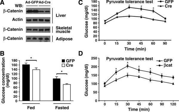
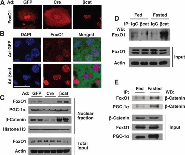
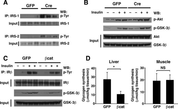
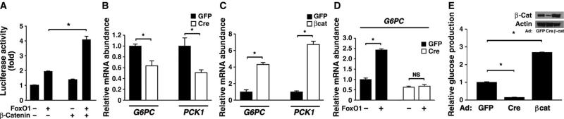
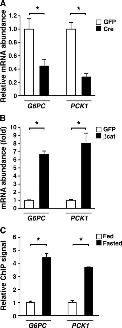
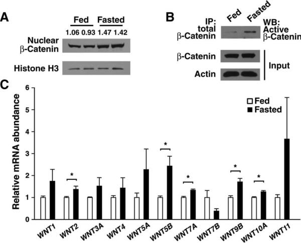
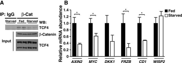
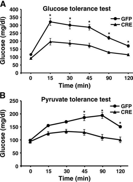
The contribution of the Wnt pathway has been extensively characterized in embryogenesis, differentiation, and stem cell biology but not in mammalian metabolism. Here, using in vivo gain- and loss-of-function models, we demonstrate an important role for Wnt signaling in hepatic metabolism. In particular, β-catenin, the downstream mediator of canonical Wnt signaling, altered serum glucose concentrations and regulated hepatic glucose production. β-Catenin also modulated hepatic insulin signaling. Furthermore, β-catenin interacted with the transcription factor FoxO1 in livers from mice under starved conditions. The interaction of FoxO1 with β-catenin regulated the transcriptional activation of the genes encoding glucose-6-phosphatase (G6Pase) and phosphoenolpyruvate carboxykinase (PEPCK), the two rate-limiting enzymes in hepatic gluconeogenesis. Moreover, starvation induced the hepatic expression of mRNAs encoding different Wnt isoforms. In addition, nutrient deprivation appeared to favor the association of β-catenin with FoxO family members, rather than with members of the T cell factor of transcriptional activators. Notably, in a model of diet-induced obesity, hepatic deletion of β-catenin improved overall metabolic homeostasis. These observations implicate Wnt signaling in the modulation of hepatic metabolism and raise the possibility that Wnt signaling may play a similar role in the metabolic regulation of other tissues.


Figures

Fig. 1
Fig. 1
Regulation of hepatic glucose metabolism by β-catenin. (A) Western blot analysis for β-catenin abundance from various tissues 7 days after tail vein infusion of adenoviruses encoding either Cre recombinase or a GFP control. Shown are protein lysates obtained from two representative mice for each condition from one of three similar experiments. (B) Serum glucose concentrations in β-catenin floxed mice previously infected with an adenovirus encoding GFP or Cre recombinase. Fasting was for 5 hours. n = 8 mice per condition. *P < 0.05, Student's t test. (C) Hepatic gluconeogenesis as assessed by pyruvate tolerance tests (PTTs) in control or β-catenin– deleted (Cre) mice. n = 8 mice per group. *P < 0.05, ANOVA with Bonferroni correction. (D) PTT in control GFP-infected mice or in mice overexpres-sing β-catenin. n = 4 mice per group. *P < 0.05, ANOVA with Bonferroni correction.
Fig. 2
Fig. 2
Interaction of FoxO1 with β-catenin. (A) Primary hepatocytes isolated from β-catenin floxed mice were infected with the indicated adenoviruses, and FoxO1 subcellular localization was subsequently assessed by indirect immunofluorescence. (B) In vivo localization of FoxO1 was determined in mice previously infected with the indicated adenoviruses and starved overnight. The Ad-βcat vector is a bicistronic construct that also encodes GFP. (C) Western blot analysis for the abundance of nuclear FoxO1, PGC-1α, and β-catenin. Nuclear fractions were prepared from mice previously infected with the indicated adenoviruses. Histone H3 was used as a loading control for nuclear proteins and actin as a control for the total hepatic lysate. (D) Lysates were prepared from whole livers of either fed mice or animals starved for 18 hours. Equal amounts of protein were immunoprecipitated (IP) with an antibody directed against β-catenin or with a control IgG serum, and the amount of coprecipitating FoxO1 was assessed by Western blot (WB) analysis. (E) Nutrient-sensitive protein interactions in fed and fasted livers observed between β-catenin and both FoxO1 and PGC-1a. Scale bar in immunohistochemical panels, 20 μm. All Western blots are representative of experiments that were performed at least three times.
Fig. 3
Fig. 3
β-Catenin modulates in vivo hepatic insulin signaling. (A) Assessment of IRS-1 and IRS-2 abundance and tyrosine phosphorylation in GFP control animals or Ad-Cre–infected mice (n = 3). Deletion of β-catenin results in higher basal tyrosine phosphorylation of IRS-1 and IRS-2. (B) Analysis of phosphorylation of Akt Ser473 and GSK-3β Ser9 under basal conditions or 5 min after insulin injection. Signaling was assessed in control GFP mice (n = 4 mice; 2 insulin-stimulated) or a similar number of Ad-Cre–infected animals. (C) Influence of β-catenin overexpression on in vivo insulin signaling. Insulin-stimulated tyrosine phosphorylation of the insulin receptor β chain (IRβ) and serine phosphorylation of GSK-3β were reduced in mice that overexpressed β-catenin. (D) In vivo assessment of de novo glycogen incorporation in control (GFP) or β-catenin–overexpressing mice. Both hepatic and skeletal muscle glycogen syntheses were assessed. n = 8 mice per group. All Western blots are representative of experiments that were performed at least three times. *P < 0.05, Student's t test. NS, not significant.
Fig. 4
Fig. 4
β-Catenin regulates in vitro transcription of two hepatic gluconeogenic enzymes and glucose production. (A) The hepatoma cell line Hepa1-6 was transiently transfected with expression plasmids encoding FoxO1, β-catenin, or empty vector (−). The activity of a FoxO-dependent luciferase promoter construct was normalized to an internal Renilla control reporter. Shown is one experiment performed in triplicate that is representative of at least three similar experiments. (B) Hepatocytes isolated from β-catenin flox/flox mice were infected with Ad-GFP or Ad-Cre, and the expression of G6PC and PCK1 was determined. n = 6 mice per group. (C) Primary mouse hepatocytes were infected with Ad-GFP or Ad-βcat, and G6PC and PCK1 expression was assessed. n = 6 mice per group. (D) Primary hepatocytes were isolated from β-catenin flox/flox mice and subsequently infected with Ad-Cre or the control Ad-GFP. Hepatocytes were then reinfected with an adenovirus encoding FoxO1, and G6PC gene expression was assessed. The deletion of β-catenin reduces FoxO1-stimulated gene expression. n = 6 mice per group. (E) Hepatic glucose production from primary hepatocytes obtained from β-catenin flox/flox mice that were infected with adenoviruses encoding GFP, Cre recombinase, or β-catenin. n = 6 mice per group. Inset shows corresponding β-catenin protein abundance.
Fig. 5
Fig. 5
In vivo regulation of the gluconeogenic program by bcatenin. (A) Relative expression of G6PC and PCK1 after in vivo deletion of hepatic β-catenin. n = 3; triplicate determinations from three animals in each group. (B) Expression of G6PC and PCK1 3 days after delivery of either a control or a β-catenin–expressing adenovirus. n = 4; triplicate determinations from four mice per group. (C) ChIP assay demonstrating increased in vivo β-catenin binding to the G6PC and PCK1 promoter after mice were fasted for 18 hours. n = 6 mice per condition. *P < 0.05, Student's t test.
Fig. 6
Fig. 6
Wnt activity is regulated by nutrient status. (A) β-Catenin abundance observed in hepatic nuclei isolated from pairs of fed or starved mice. Numerical values represent the arbitrary ratio of nuclear β-catenin to histone H3 abundance.(B) Abundance of active β-catenin in fed and fasting livers. Total β-catenin was immunoprecipitated from equal amounts (2 mg) of hepatic protein lysate and then assessed by Western blot (WB) analysis using an antibody that recognizes the active (hypophosphorylated) form of β-catenin. All Western blots are representative of experiments that were performed at least three times. (C) In vivo expression of various Wnt ligands under fed and starved conditions. n = 3 to 4 mice per condition, each assessed in triplicate. *P < 0.05, Student's t test.
Fig. 7
Fig. 7
Starvation alters β-catenin binding to TCF4. (A) Hepatic lysates were prepared from fed or starved mice to assess the interaction of β-catenin with TCF4. Lysates were immunoprecipitated with a β-catenin antibody or an IgG control serum. Interaction of β-catenin with TCF4 was determined by immunoprecipitation of β-catenin followed by Western blot analysis of TCF4. Although the abundance of β-catenin and TCF4 was not altered by starvation, the interaction of β-catenin with TCF4 was reduced under starved conditions. TCF4 appeared as a long and a short isoform in hepatic lysates, of which only the long form appeared to interact strongly with β-catenin. (B) Analysis of known Wnt target genes under fed or starved conditions. Consistent with the decline in TCF4 binding, most previously identified β-catenin/TCF4 targets decreased in expression under starved conditions. n = 4 to 6 determinations per gene. *P < 0.05, Student's t test.
Fig. 8
Fig. 8
Deletion of β-catenin improves glucose homeostasis in a model of diet-induced obesity. (A) Glucose tolerance test of β-catenin floxed mice on a high-fat diet randomized to tail vein injection of adenoviruses encoding Cre recombinase or GFP. (B) PTT of control mice or mice with Cre recombinase–mediated deletion of β-catenin. n = 6 mice per group. *P < 0.05 by ANOVA with Bonferroni correction.

References

    1. Petersen CP, Reddien PW. Wnt signaling and the polarity of the primary body axis. Cell. 2009;139:1056–1068. - PubMed
    1. van Amerongen R, Nusse R. Towards an integrated view of Wnt signaling in development. Development. 2009;136:3205–3214. - PubMed
    1. Reya T, Clevers H. Wnt signalling in stem cells and cancer. Nature. 2005;434:843–850. - PubMed
    1. Angers S, Moon RT. Proximal events in Wnt signal transduction. Nat. Rev. Mol. Cell Biol. 2009;10:468–477. - PubMed
    1. MacDonald BT, Tamai K, He X. Wnt/β-catenin signaling: Components, mechanisms, and diseases. Dev. Cell. 2009;17:9–26. - PMC - PubMed

MeSH terms