

Mutations in the human SC4MOL gene encoding a methyl sterol oxidase cause psoriasiform dermatitis, microcephaly, and developmental delay
- PMID: 21285510
- PMCID: PMC3049385
- DOI: 10.1172/JCI42650
Mutations in the human SC4MOL gene encoding a methyl sterol oxidase cause psoriasiform dermatitis, microcephaly, and developmental delay
Abstract
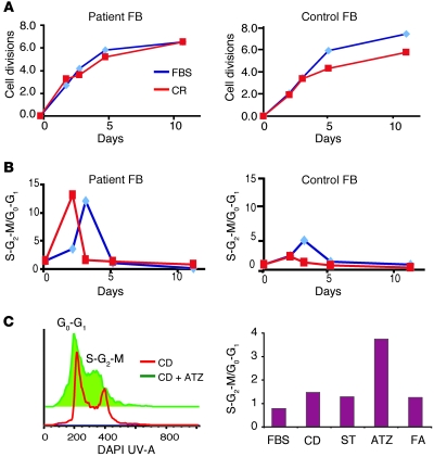
Defects in cholesterol synthesis result in a wide variety of symptoms, from neonatal lethality to the relatively mild dysmorphic features and developmental delay found in individuals with Smith-Lemli-Opitz syndrome. We report here the identification of mutations in sterol-C4-methyl oxidase–like gene (SC4MOL) as the cause of an autosomal recessive syndrome in a human patient with psoriasiform dermatitis, arthralgias, congenital cataracts, microcephaly, and developmental delay. This gene encodes a sterol-C4-methyl oxidase (SMO), which catalyzes demethylation of C4-methylsterols in the cholesterol synthesis pathway. C4-Methylsterols are meiosis-activating sterols (MASs). They exist at high concentrations in the testis and ovary and play roles in meiosis activation. In this study, we found that an accumulation of MASs in the patient led to cell overproliferation in both skin and blood. SMO deficiency also substantially altered immunocyte phenotype and in vitro function. MASs serve as ligands for liver X receptors α and β(LXRα and LXRβ), which are important in regulating not only lipid transport in the epidermis, but also innate and adaptive immunity. Deficiency of SMO represents a biochemical defect in the cholesterol synthesis pathway, the clinical spectrum of which remains to be defined.
Figures


Similar articles
-
A rare case of sterol-C4-methyl oxidase deficiency in a young Italian male: Biochemical and molecular characterization.Mol Genet Metab. 2017 Aug;121(4):329-335. doi: 10.1016/j.ymgme.2017.06.013. Epub 2017 Jun 27. Mol Genet Metab. 2017. PMID: 28673550
-
The role of sterol-C4-methyl oxidase in epidermal biology.Biochim Biophys Acta. 2014 Mar;1841(3):331-5. doi: 10.1016/j.bbalip.2013.10.009. Epub 2013 Oct 18. Biochim Biophys Acta. 2014. PMID: 24144731 Free PMC article. Review.
-
New Homozygous Missense MSMO1 Mutation in Two Siblings with SC4MOL Deficiency Presenting with Psoriasiform Dermatitis.Cytogenet Genome Res. 2020;160(9):523-530. doi: 10.1159/000511126. Epub 2020 Nov 6. Cytogenet Genome Res. 2020. PMID: 33161406
-
MSMO1 deficiency: a potentially partially treatable, ultrarare neurodevelopmental disorder with psoriasiform dermatitis, alopecia and polydactyly.Clin Dysmorphol. 2023 Jul 1;32(3):97-105. doi: 10.1097/MCD.0000000000000461. Epub 2023 May 8. Clin Dysmorphol. 2023. PMID: 37195326
-
Exome sequencing identifies a de novo mutation of CTNNB1 gene in a patient mainly presented with retinal detachment, lens and vitreous opacities, microcephaly, and developmental delay: Case report and literature review.Medicine (Baltimore). 2017 May;96(20):e6914. doi: 10.1097/MD.0000000000006914. Medicine (Baltimore). 2017. PMID: 28514307 Free PMC article. Review.
Cited by
-
Differential Methylation of H3K79 Reveals DOT1L Target Genes and Function in the Cerebellum In Vivo.Mol Neurobiol. 2019 Jun;56(6):4273-4287. doi: 10.1007/s12035-018-1377-1. Epub 2018 Oct 10. Mol Neurobiol. 2019. PMID: 30302725 Free PMC article.
-
CYP5122A1 encodes an essential sterol C4-methyl oxidase in Leishmania donovani and determines the antileishmanial activity of antifungal azoles.Nat Commun. 2024 Oct 31;15(1):9409. doi: 10.1038/s41467-024-53790-5. Nat Commun. 2024. PMID: 39482311 Free PMC article.
-
Qki activates Srebp2-mediated cholesterol biosynthesis for maintenance of eye lens transparency.Nat Commun. 2021 May 21;12(1):3005. doi: 10.1038/s41467-021-22782-0. Nat Commun. 2021. PMID: 34021134 Free PMC article.
-
MicroRNA-223 coordinates cholesterol homeostasis.Proc Natl Acad Sci U S A. 2014 Oct 7;111(40):14518-23. doi: 10.1073/pnas.1215767111. Epub 2014 Sep 22. Proc Natl Acad Sci U S A. 2014. PMID: 25246565 Free PMC article.
-
Pervasive inflammatory activation in patients with deficiency in very-long-chain acyl-coA dehydrogenase (VLCADD).Clin Transl Immunology. 2021 Jun 27;10(6):e1304. doi: 10.1002/cti2.1304. eCollection 2021. Clin Transl Immunology. 2021. PMID: 34194748 Free PMC article.
References
-
- Herman GE. Disorders of cholesterol biosynthesis: prototypic metabolic malformation syndromes. Hum Mol Genet. 2003;12 spec no. 1:R75–R88. - PubMed
Publication types
MeSH terms
Substances
Grants and funding
LinkOut - more resources
Full Text Sources
Other Literature Sources
Medical
Molecular Biology Databases
Miscellaneous
