. 2011 Apr;19(4):751-9.
doi: 10.1038/mt.2010.313. Epub 2011 Feb 1.

Improving adoptive T cell therapy by targeting and controlling IL-12 expression to the tumor environment

Affiliations

Improving adoptive T cell therapy by targeting and controlling IL-12 expression to the tumor environment

Ling Zhang et al. Mol Ther. 2011 Apr.

Abstract

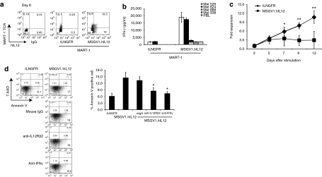
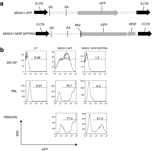
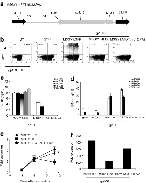
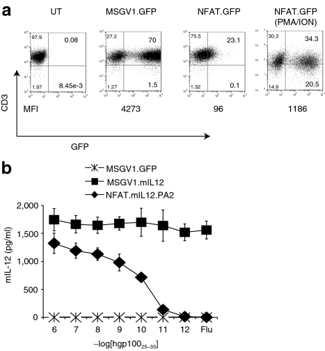
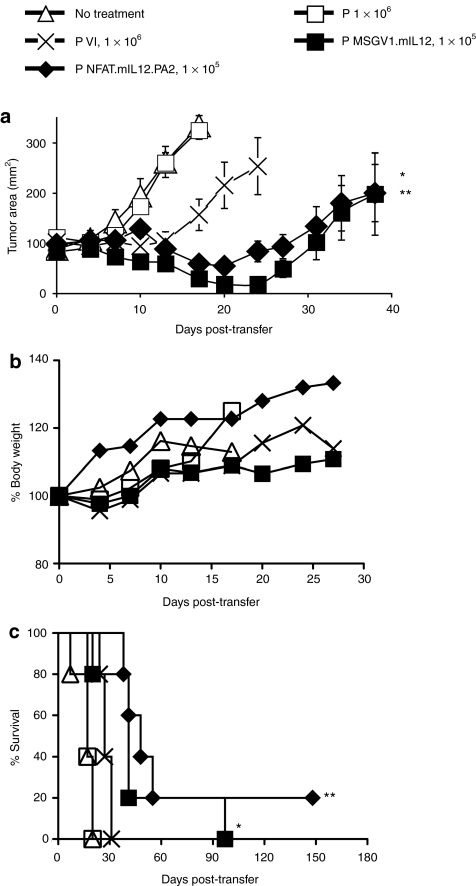
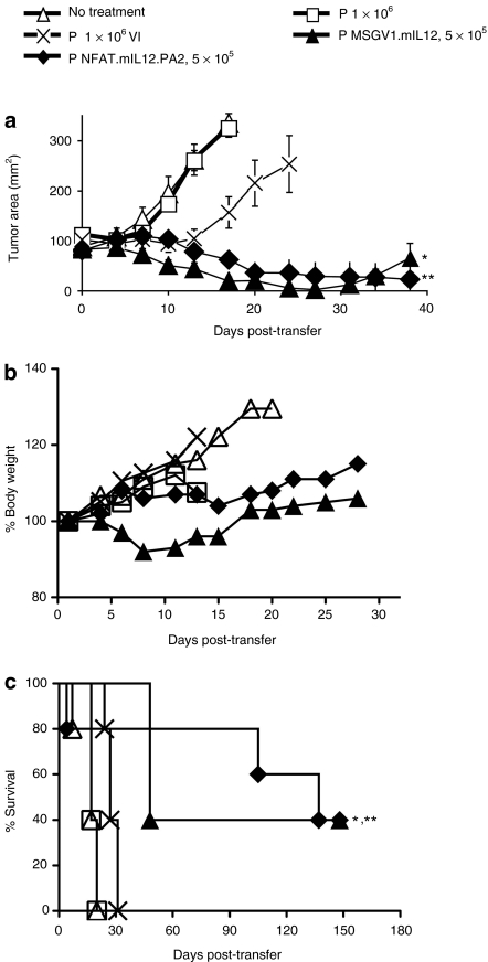
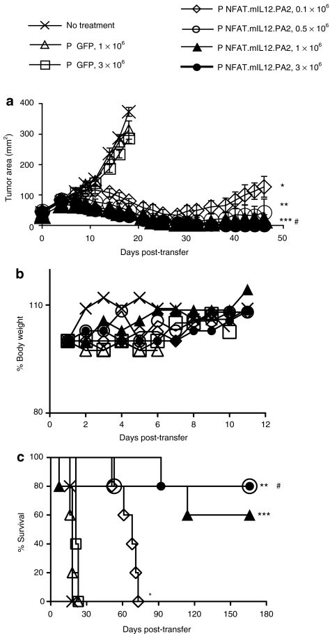
Interleukin-12 (IL-12) is an important immunostimulatory cytokine, yet its clinical application has been limited by the systemic toxicity associated with its administration. In this work, we developed a strategy to selectively deliver IL-12 to the tumor environment using genetically engineered lymphocytes. However, peripheral blood lymphocytes (PBLs) transduced with a γ-retroviral vector, which constitutively expressed IL-12, failed to expand in culture due to apoptosis. To circumvent this problem, a vector was designed where IL-12 expression was directed by a composite promoter-containing binding motifs for nuclear factor of activated T-cells (NFAT.hIL12.PA2). The NFAT-responsive promoter was activated to drive IL-12 expression upon the recognition of tumor-specific antigen mediated by a T cell receptor (TCR) that was engineered into the same lymphocytes. We tested the efficacy of the inducible IL-12 vector in vivo in a murine melanoma model. Adoptive transfer of pmel-1 T cells genetically engineered with NFAT-murineIL12 (NFAT.mIL12.PA2) significantly enhanced regression of large established B16 melanoma. Notably, this targeted and controlled IL-12 treatment was without toxicity. Taken together, our results suggest that using the NFAT.hIL12.PA2 vector might be a promising approach to enhance adoptive cancer immunotherapy.


Figures

Figure 1
Figure 1
Constitutive expression of bioactive interleukin-12 (IL-12) inhibited the proliferation of transduced peripheral blood lymphocytes (PBLs) and was associated with increased apoptosis. (a) Human PBLs were cotransduced with MART-1 T cell receptor (TCR) and MSGV1.hIL12 vector or control truncated low-affinity nerve growth factor receptor (tLNGFR) vector. Expression of MART-1 and IL-12 were measured by fluorescence-activated cell sorter (FACS) using mouse antihuman Vβ12-phycoerythrin (PE) and IL-12-fluorescein isothiocyanate (FITC) antibody on day 6 after transduction. (b) The double-transduced PBLs were cocultured with melanoma lines: mel526 (HLA-A2+/MART-1+), mel624 (HLA-A2+/MART-1+), mel888 (HLA-A2/MART-1+), mel938 (HLA-A2/MART-1+). Concentration of interferon (IFN)-γ in the culture was measured using IFN-γ enzyme-linked immunosorbent assay (ELISA) (shown are the mean values of triplicate determinations, ±SD). (c) PBLs from three donors were transduced with tLNGFR or MSGV1.hIL12 to evaluate in vitro cell expansion. Viable cells were enumerated every 3–5 days by Trypan blue exclusion (n = 3; *P < 0.05; **P < 0.001). (d) Left panel: IL-12-induced apoptosis in PBLs was partially blocked by anti-IFN-γ or anti-IL12Rβ2 antibodies. On day 2 after transduction, the transduced cells were treated with 50 µg/ml of indicated antibodies. Three days later, the cells were stained with Annexin V-phycoerythrin (PE) and 7-aminoactinomycin D (7-AAD) and analyzed by FACS. For each plot, the upper right sextant contains necrotic cells, the middle right contains late apoptotic cells, and the lower right contains early apoptotic cells. (d) Right panel: quantification of the percentage of Annexin V positive staining cells in each treatment condition (n = 3, shown are the mean values of triplicate determinations, ±SD, *P < 0.05 compared to mIgG).
Figure 2
Figure 2
Green fluorescent protein (GFP) expression driven by an nuclear factor of activated T-cells (NFAT) responsive promoter in transduced peripheral blood lymphocytes (PBLs) was triggered by T cell activation. (a) Schematic representation of retroviral vectors: MSGV1.GFP and MSGV1.NFAT.GFP.PA2 GFP, enhanced green fluorescence protein; LTR, long-terminal repeat; NFAT, composite NFAT-responsive promoter element; PA2, polyadenylation signal; SA, splice acceptor; SD, splice donor. (b) Vectors were transfected 293GP cell using lipofectamine 2000 for transient virus production (top histograms). PBLs were transduced with individual vectors as described in the methods. Two days later, the cells were left untreated (middle histograms) or activated with phorbol myristate acetate (PMA)/ionomycin overnight (bottom histograms). Expression of GFP in 293GP cells and PBLs was measured by fluorescence-activated cell sorter (FACS), the percent positive cells was as shown. UT, untransduced.
Figure 3
Figure 3
Nuclear factor of activated T-cells (NFAT) responsive promoter-directed expression of interleukin-12 (IL-12) triggered by T cell receptor (TCR) engagement. (a) Diagram of γ-retroviral vector-containing NFAT.hIL12.PA2 expression cassette. hscIL12, human single chain IL-12 protein; LTR, long-terminal repeat; NFAT, composite NFAT-responsive promoter element; PA2, polyadenylation signal; SA, splice acceptor; SD, splice donor. (b) OKT3 stimulated peripheral blood lymphocytes (PBLs) were transduced with a retroviral vector encoding TCR recognizing melanoma tumor antigen gp100 on day 2. The next day, cells were subject to a second transduction with vectors, MSGV1.GFP, MSGV1.hIL12 or MSGV1.NFAT.hIL12.PA2. On day 7, cells were analyzed by flow cytometry to detect expression of TCR and green fluorescent protein (GFP). UT, untransduced. (c,d) Double-transduced cells were cocultured with melanoma lines: mel 624 (HLA-A2+/gp100+), mel 526 (HLA-A2+/gp100+), mel 888 (HLA-A2/gp100+), or mel 938 (HLA-A2/gp100+). The concentration of (c) IL-12 and (d) IFN-γ in the coculture media was measured by enzyme-linked immunosorbent assay (ELISA) (shown are the mean values of triplicate determinations, ±SD). (e) The proliferation of transduced cells for multiple cultures (n = 4) was measured by counting viable cells every 3–5 days after Trypan blue exclusion (*P < 0.001 for the NFAT-IL12 vector compared to the constitutive IL-12 vector at day 11). (f) On day 9, the transduced cells were subject to a rapid expansion by culture with allogeneic peripheral blood mononuclear cell (PBMC) feeder cells (treated by 4,000 rad irradiation) and 6,000 IU/ml IL-2. The viable cells were enumerated 11 days later. The data are the average fold expansion from two different donors. The data in figure bd are representative of one of four experiments.
Figure 4
Figure 4
Human nuclear factor of activated T-cells (NFAT) responsive promoter induced gene expression in murine pmel-1 T cells after stimulation. (a) NFAT-responsive promoter drives green fluorescent protein (GFP) expression in pmel-1 cells. Pmel splenocytes were stimulated with 1 µmol/l hgp10025–33 peptide and transduced with retroviral vector MSGV1.GFP or NFAT.GFP.PA2. Two days after transduction, the cells were stimulated with phorbol myristate acetate (PMA)/ionomycin overnight and analyzed by flow cytometry for CD3 and GFP. The percentage of positive cells in each quadrant was as shown, with the mean fluorescence intensity (MFI) indicated below. UT, untransduced. (b) Murine IL-12 was induced in gene engineered pmel-1 T cells. Pmel-1 splenocytes were transduced with MSGV1.mIL12, NFAT.mIL12.PA2, or MSGV1.GFP after stimulation by the hgp10025–33 peptide. The cells were then activated by cocultured with C57BL/6 splenocytes pulsed with hgp10025–33 peptide at various concentration for 16 hours (Flu, control influenza virus peptide). Production of IL-12 in the coculture media was detected by enzyme-linked immunosorbent assay (ELISA) (shown are the mean values of triplicate determinations, ±SD).
Figure 5
Figure 5
Adoptive transfer of Pmel-1 T cells engineered with mIL-12 caused regression of large established B16 melanoma without the need for interleukin-2 (IL-2) administration and vaccination. (a) Pmel-1 T cells were transduced with mIL-12 viral vectors, including MSGV1.mIL12*, MSGV1.NFAT.mIL12.PA2**. Tumor-bearing C57BL/6 mice (n = 5) were adoptive transferred with 1 × 10⁶ native pmel-1 T cells only (P only) or 1 × 10⁶ native pmel-1 T cells combined with IL-2 600,000 IU daily for 3 days and rVVhgp100 vaccine (PVI). 1 × 10⁵ pmel-1 T cells engineered with mIL-12 vectors were transferred without IL-2 and vaccine. All the mice received 5 Gy lymphodepleting irradiation before infusion. Tumor sizes were assessed with serial measurements. Error bars represent SE of the mean (*,**P < 0.01 compared with PVI). (b) Body weight of each group was measure at different days (baseline = 100%). (c) The survival of tumor-bearing mice that received cell transfer was determined as shown (*,**P < 0.05 compared with PVI). The data represents one of two independent experiments (n = 5 animals per group).
Figure 6
Figure 6
Adoptive transfer of increased numbers of interleukin-12 (IL-12) modified cells improved the treatment efficacy but total body weight loss was observed using the MSGV1.mIL12 vector. (a) Tumor-bearing C57BL/6 mice (n = 5) were adoptive transferred with 1 × 10⁶ native pmel-1 T cells only (P only) or 1 × 10⁶ native pmel-1 T cells combined with IL-2 600,000 IU daily for 3 days and rVVhgp100 vaccine (PVI). 5 × 10⁵ pmel-1 T cells engineered with mIL-12 vectors (MSGV1.mIL12* or NFAT.mIL12.PA2**) were transferred without IL-2 and vaccine. All the mice received 5 Gy lymphodepletion irradiation before infusion. Tumor sizes were assessed with serial measurements. Error bars represent SE of the mean (*,**P < 0.005 compared with PVI). (b) Body weight of each group was measure at different days (baseline = 100%). (c) The survival of tumor-bearing mice that received cell transfer was determined as shown (*,**P < 0.05 compared with PVI).
Figure 7
Figure 7
Adoptive transfer of large numbers of Pmel-1 T cells engineered with NFAT.IL12.PA2 vector-enhanced tumor therapeutic efficacy without observed toxicity. (a) Antitumor treatment by transferring of 0.1 × 10⁶ *, 0.5 × 10⁶ **, 1 × 10⁶ ***, and 3 × 10⁶ # pmel-1 T cells engineered with NFAT.mIL12.PA2 into B16 tumor-bearing mice (n = 5 animals per group, green fluorescent protein (GFP) transduced pmel T cells used as treatment control). All the mice received 5 Gy lymphodepletion irradiation before infusion, there was no vaccine or IL-2 administration. Tumor sizes were assessed with serial measurements. Error bars represent the SE of the mean (*,**,***,#P < 0.01 compared with pmel-T cell transduced with GFP). (b) Body weight of each group was measure at different days (baseline = 100%). (c) The survival of tumor-bearing mice that received pmel-1 T cells engineered with the NFAT.IL12.PA2 vectors or GFP was determined as shown (*,**,***,#P < 0.05 compared with pmel-T cell transduced with GFP).

References

    1. Kobayashi M, Fitz L, Ryan M, Hewick RM, Clark SC, Chan S.et al. (1989Identification and purification of natural killer cell stimulatory factor (NKSF), a cytokine with multiple biologic effects on human lymphocytes J Exp Med 170827–845. - PMC - PubMed
    1. Gubler U, Chua AO, Schoenhaut DS, Dwyer CM, McComas W, Motyka R.et al. (1991Coexpression of two distinct genes is required to generate secreted bioactive cytotoxic lymphocyte maturation factor Proc Natl Acad Sci USA 884143–4147. - PMC - PubMed
    1. Gately MK. Interleukin-12: a recently discovered cytokine with potential for enhancing cell-mediated immune responses to tumors. Cancer Invest. 1993;11:500–506. - PubMed
    1. Mehrotra PT, Wu D, Crim JA, Mostowski HS., and, Siegel JP. Effects of IL-12 on the generation of cytotoxic activity in human CD8+ T lymphocytes. J Immunol. 1993;151:2444–2452. - PubMed
    1. Manetti R, Parronchi P, Giudizi MG, Piccinni MP, Maggi E, Trinchieri G.et al. (1993Natural killer cell stimulatory factor (interleukin 12 [IL-12]) induces T helper type 1 (Th1)-specific immune responses and inhibits the development of IL-4-producing Th cells J Exp Med 1771199–1204. - PMC - PubMed

Publication types

MeSH terms

LinkOut - more resources