Cervical radiculopathy caused by vertebral artery loop formation : a case report and review of the literature
- PMID: 21286489
- PMCID: PMC3030092
- DOI: 10.3340/jkns.2010.48.5.465
Cervical radiculopathy caused by vertebral artery loop formation : a case report and review of the literature
Abstract
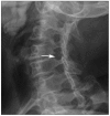
Vertebral artery loop formation causing encroachment on cervical neural foramen and canal is a rare cause of cervical radiculopathy. We report a case of 61-year-old woman with vertebral artery loop formation who presented with right shoulder pain radiating to her arm for 2 years. Plain radiograph and computed tomography scan revealed widening of the right intervertebral foramen at the C5-6 level. Magnetic resonance imaging and angiogram confirmed the vertebral artery loop formation compressing the right C6 nerve root. We had considered microdecompressive surgery, but the patient's symptoms resolved after conservative management. Clinician should keep in mind that vertebral artery loop formation is one of important causes of cervical radiculopathy. Vertebral artery should be visualized using magnetic resonance angiography in suspected case.
Keywords: Cervical radiculopathy; Magnetic resonance angiography; Vascular anomaly; Vascular compression; Vertebral artery; Vertebral artery loop.
Figures


References
-
- Anderson RE, Shealy CN. Cervical pedicle erosion and rootlet compression caused by a tortuous vertebral artery. Radiology. 1970;96:537–538. - PubMed
-
- Barratt JG. Enlargement of cervical intervertebral foramina by coiling of the vertebral artery. Australas Radiol. 1974;18:171–174. - PubMed
-
- Bruneau M, Cornelius JF, Marneffe V, Triffaux M, George B. Anatomical variations of the V2 segment of the vertebral artery. Neurosurgery. 2006;59:ONS20–ONS24. discussion ONS20-ONS24. - PubMed
-
- Burnett KR, Staple TW. Case report 132. Tortuous vertebral artery causing erosive defect of C2. Skeletal Radiol. 1981;6:51–53. - PubMed
-
- Cooper DF. Bone erosion of the cervical vertebrae secondary to tortuosity of the vertebral artery : case report. J Neurosurg. 1980;53:106–108. - PubMed
Publication types
LinkOut - more resources
Full Text Sources
Research Materials
Miscellaneous

