. 2011 Sep;469(9):2558-67.
doi: 10.1007/s11999-011-1775-4.

Bony increased-offset reversed shoulder arthroplasty: minimizing scapular impingement while maximizing glenoid fixation

Affiliations

Bony increased-offset reversed shoulder arthroplasty: minimizing scapular impingement while maximizing glenoid fixation

Pascal Boileau et al. Clin Orthop Relat Res. 2011 Sep.

Abstract

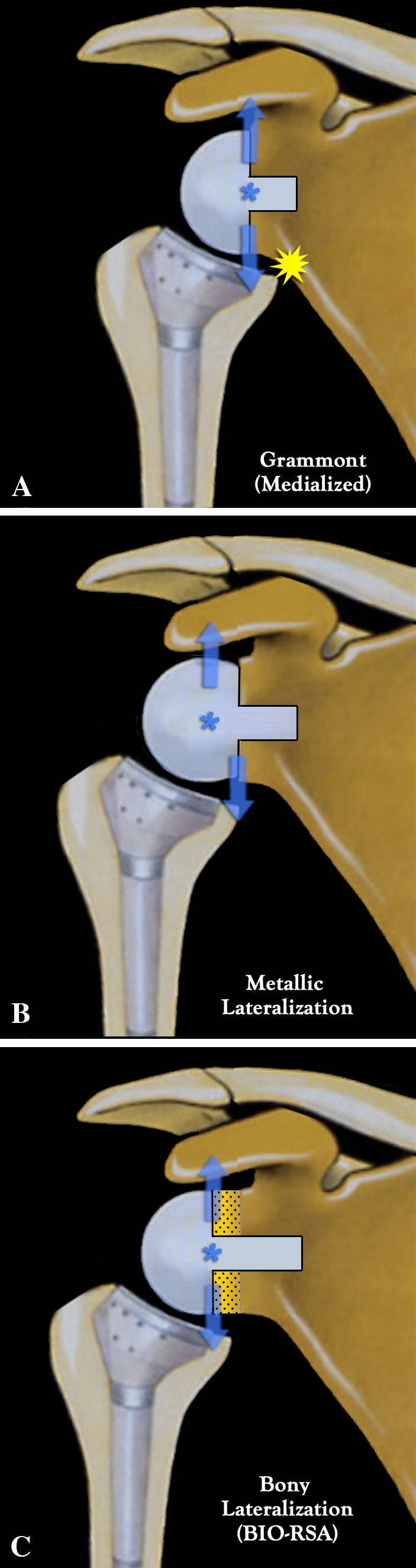
Background: Scapular notching, prosthetic instability, limited shoulder rotation and loss of shoulder contour are associated with conventional medialized design reverse shoulder arthroplasty. Prosthetic (ie, metallic) lateralization increases torque at the baseplate-glenoid interface potentially leading to failure.

Questions/purposes: We asked whether bony lateralization of reverse shoulder arthroplasty would avoid the problems caused by humeral medialization without increasing torque or shear force applied to the glenoid component.

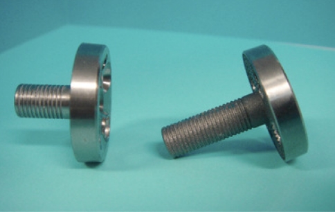
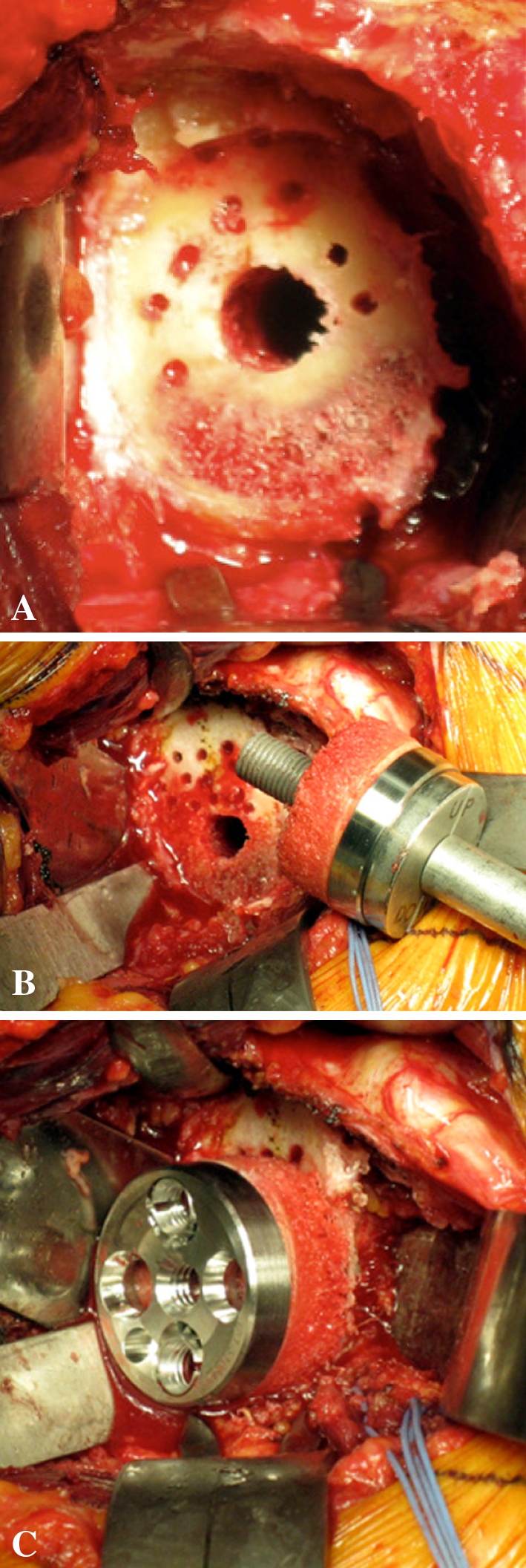
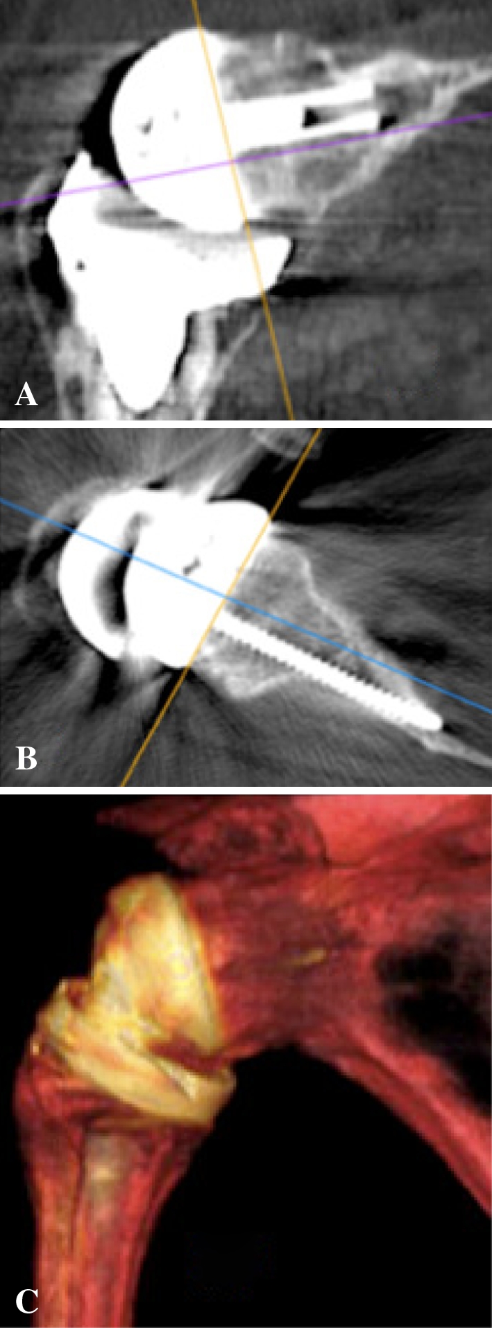
Patients and methods: We prospectively followed 42 patients with rotator cuff deficiency treated with bony increased-offset reverse shoulder arthroplasty. A cylinder of autologous cancellous bone graft, harvested from the humeral head, was placed between the reamed glenoid surface and baseplate. Graft and baseplate fixation was achieved using a lengthened central peg (25 mm) and four screws. Patients underwent clinical, radiographic, and CT assessment at a minimum of 2 years after surgery.

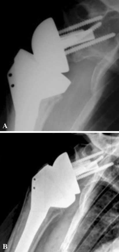
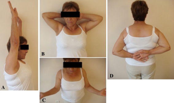
Results: The humeral graft incorporated completely in 98% of cases (41 of 42) and partially in one. At a mean of 28 months postoperatively, no graft resorption, glenoid loosening, or postoperative instability was observed. Inferior scapular notching occurred in 19% (eight of 42). The absolute Constant-Murley score improved from 31 to 67. Thirty-six patients (86%) were able to internally rotate sufficiently to reach their back over the sacrum.

Conclusions: Grafting of the glenoid surface during reverse shoulder arthroplasty effectively creates a long-necked scapula, providing the benefits of lateralization. Bony increased-offset reverse shoulder arthroplasty is associated with low rates of inferior scapular notching, improved shoulder rotation, no prosthetic instability and improved shoulder contour. In contrast to metallic lateralization, bony lateralization has the advantage of maintaining the prosthetic center of rotation at the prosthesis-bone interface, thus minimizing torque on the glenoid component.

Level of evidence: Level IV, therapeutic study. See Guidelines for Authors for a complete description of levels of evidence.


Figures

Fig. 1A–C
Fig. 1A–C
Diagrams show medialization versus lateralization in RSA. (A) Medialized (Grammont) RSA (hemisphere) places the center of rotation at the bone-prosthesis interface. Deltoid force applied to the center of rotation does not develop any torque because there is no lever arm, but there is a risk of scapular notching. (B) Metallic lateralized RSA (two-thirds of a sphere) reduces the risk of scapular notching but at the price of creating a lever arm because a lateralized center of rotation produces shear forces detrimental to glenoid fixation. (C) BIO-RSA reduces the risk of scapular notching (due to the lateralization), while maximizing glenoid fixation (because the center of rotation remains at the bone-prosthesis interface and there is no lever arm).
Fig. 2
Fig. 2
A photograph shows a standard baseplate with a 15-mm-long central peg (left) and a BIO-RSA baseplate with a lengthened, 25-mm-long peg (right) used for primary fixation of the bone graft in the native glenoid vault.
Fig. 3A–E
Fig. 3A–E
Photographs illustrate the cancellous bone graft being harvested from the humerus. (A) A bell saw is used to create a cylinder of cancellous bone. (B) The cylinder of cancellous bone is shown. (C) A cutting guide is inserted to harvest the desired thickness of bone graft. The disc of cancellous bone graft is (D) retrieved from the cutting guide and (E) inserted along the lengthened central peg of the baseplate.
Fig. 4A–C
Fig. 4A–C
Photographs illustrate glenoid preparation. (A) The glenoid surface must be reamed until the subchondral plate is reached (this represents an approximate 5-mm deep reaming) and with some inferior tilt. In addition to glenoid reaming, small drill holes (2.5 mm) are made at the periphery of the glenoid to obtain a complete bleeding bone surface and the central hole is overdrilled with an 8-mm drill bit. (B) The baseplate, with the disc of cancellous bone graft inserted along the lengthened central peg. (C) It is impacted until it reaches the cancellous glenoid surface and then fixed with four (two compressive and two locking) screws.
Fig. 5A–B
Fig. 5A–B
(A) An AP radiograph performed 3 months after surgery demonstrates complete bone graft healing. (B) No bone graft resorption or lysis and no scapular notching are observed at 36 months’ followup. Note the low and inferiorly tilted positioning of the glenoid implant in addition to its lateralization.
Fig. 6A–C
Fig. 6A–C
CT scans at 38 months’ followup show bone graft healing in both (A) coronal and (B) axial planes. (C) A three-dimensional reconstruction demonstrates the lengthened scapular neck obtained after bone graft healing. The bony lateralization offers the advantage of keeping the humeral cup away from the pillar of the scapula, thus decreasing the risk of impingement while keeping the center of rotation within the bone.
Fig. 7A–D
Fig. 7A–D
Photographs of a 70-year-old patient 29 months after BIO-RSA performed on the right shoulder show (A) active anterior elevation of 170°, (B) elevation and external rotation, (C) external rotation of 45°, and (D) internal rotation to L1. Notice the shoulder contour which is similar to the contralateral side.

Comment in

References

    1. Abrassart S, Stern R, Hoffmeyer P. Arterial supply of the glenoid: an anatomic study. J Shoulder Elbow Surg. 2006;15:232–238. doi: 10.1016/j.jse.2005.07.015. - DOI - PubMed
    1. Antuna S, Sperling JW, Cofield RH. Reimplantation of a glenoid component after component removal and allograft bone grafting: a report of 3 cases. J Shoulder Elbow Surg. 2002;11:637–641. doi: 10.1067/mse.2002.126100. - DOI - PubMed
    1. Baulot E, Chabernaud D, Grammont PM. Results of Grammont’s inverted prosthesis in omarthritis associated with major cuff destruction: apropos of 16 cases [in French] Acta Orthop Belg. 1995;61(Suppl 1):112–119. - PubMed
    1. Boileau P, Chuinard C, Roussanne Y, Bicknell RT, Rochet N, Trojani C. Reverse shoulder arthroplasty combined with a modified latissimus dorsi and teres major tendon transfer for shoulder pseudoparalysis associated with dropping arm. Clin Orthop Relat Res. 2008;466:584–593. doi: 10.1007/s11999-008-0114-x. - DOI - PMC - PubMed
    1. Boileau P, Watkinson DJ, Hatzidakis AM, Balg F. Grammont reverse prosthesis: design, rationale, and biomechanics. J Shoulder Elbow Surg. 2005;14(1 Suppl):147S–161S. doi: 10.1016/j.jse.2004.10.006. - DOI - PubMed

MeSH terms