. 2011 Mar;79(3):947-64.
doi: 10.1002/prot.22936. Epub 2010 Dec 22.

Analysis of the peroxiredoxin family: using active-site structure and sequence information for global classification and residue analysis

Affiliations

Analysis of the peroxiredoxin family: using active-site structure and sequence information for global classification and residue analysis

Kimberly J Nelson et al. Proteins. 2011 Mar.

Abstract

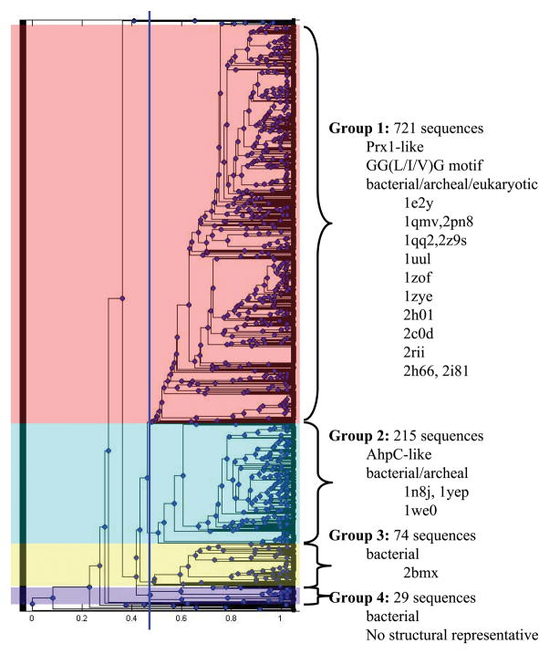
Peroxiredoxins (Prxs) are a widespread and highly expressed family of cysteine-based peroxidases that react very rapidly with H₂O₂, organic peroxides, and peroxynitrite. Correct subfamily classification has been problematic because Prx subfamilies are frequently not correlated with phylogenetic distribution and diverge in their preferred reductant, oligomerization state, and tendency toward overoxidation. We have developed a method that uses the Deacon Active Site Profiler (DASP) tool to extract functional-site profiles from structurally characterized proteins to computationally define subfamilies and to identify new Prx subfamily members from GenBank(nr). For the 58 literature-defined Prx test proteins, 57 were correctly assigned, and none were assigned to the incorrect subfamily. The >3500 putative Prx sequences identified were then used to analyze residue conservation in the active site of each Prx subfamily. Our results indicate that the existence and location of the resolving cysteine vary in some subfamilies (e.g., Prx5) to a greater degree than previously appreciated and that interactions at the A interface (common to Prx5, Tpx, and higher order AhpC/Prx1 structures) are important for stabilization of the correct active-site geometry. Interestingly, this method also allows us to further divide the AhpC/Prx1 into four groups that are correlated with functional characteristics. The DASP method provides more accurate subfamily classification than PSI-BLAST for members of the Prx family and can now readily be applied to other large protein families.


Figures

Figure 1
Figure 1. Location of residues conserved across all Prx subfamilies
The structure of the S. typhimurium AhpC (PDB identifier 1n8j) active site is shown with the location of the CP (Cys46) in yellow (the protein sequence of 1n8j contains a C46S mutation, but for simplicity it is labeled as a Cys). The adjacent monomer across the dimer interface (B interface) is in pale green and the adjacent monomer across the decamer-building interface (A interface) is in tan. In the Tpx and Prx5 subfamilies, the dimer is formed across the A-type interface. Residues conserved across the Prx family are highlighted in cyan (identified previously) and magenta (identified as part of the work described herein). Brown highlights the loop-helix region around the active site that undergoes local unfolding following oxidation to form a disulfide bond between CP and CR in both typical and atypical 2-Cys Prxs. This figure was made using Pymol (http://sourceforge.net/projects/pymol/).
Figure 2
Figure 2. Functional site signatures extracted from all Prx structures identify six Prx subfamilies
Functional site signatures were created for the active site of each Prx structure in the RCSB protein database (Jan 2008) using the DASP software package at http://dasp.deac.wfu.edu/. (A) The functional site signatures were hierarchically clustered in Matlab using the unweighted pair group method average (UPGMA) algorithm. The dendrogram shown illustrates the resulting organization of functional site signatures and does not imply any evolutionary relationship. The cluster for each Prx subfamily is highlighted and labeled with the subfamily name, taken from one or two prototypical members of that subfamily. (B) Alignment of functional site signatures for Prx proteins of known structure identifies sequence characteristics for each subfamily. Changes between upper and lower case letters across each line denote a change to the next piece of contiguous protein sequence in a signature. Residues that are conserved across all Prxs are highlighted in black and key residues used to create the functional site signatures are starred. Residues conserved across each subfamily based upon analysis of proteins in the PDB database are highlighted in gray; residues found to be conserved within each subfamily following analysis of GenBank(nr) sequences are boxed. Only one signature is shown for any protein with multiple structures in the PDB database. Any engineered mutations, oxidized forms of cysteine, or seleno-methionine residues were changed back to their wild-type residue prior to alignment. This profile was created by first aligning the signatures for each subfamily using ClustalW and then editing by hand to correctly align the key residues across all subfamilies. Because of the variability among the signatures, DASP was unable to score this complete profile.
Figure 3
Figure 3. Residue conservation and potential structural/functional role for each residue in Prx subfamily functional site profiles
The functional site signatures are shown for a representative member of each of the Prx subfamilies. The degree of conservation is shown for each residue after aligning the full sequence for all of the putative members identified from the GenBank(nr) database searches. The actual residues and numbers listed are for (A) Aeropyrum pernix BCP (2cx4), (B) Salmonella typhimurium AhpC (1yep) and Trypanosoma cruzi tryparedoxin peroxidase (1uul), (C) Homo sapiens Prx6 (1prx), (D) H. sapiens Prx5 (1hd2), and (E) Streptococcus pneumoniae Tpx (1psq). Residues with an entropy value below 0.61 (mean minus 1 standard deviation) are considered conserved (dashed line). The potential role for each conserved residue is also represented in the histogram and labeled as follows: the peroxidatic cysteine (Cp), the resolving cysteine (Cr), key residues conserved across all Prx subfamilies (▲), residues involved in forming the active site pocket of the reduced protein (§), residues found in the A-type interface (A), residues found in the B-type interface (B), residues involved in stabilizing the helix containing the CP (●), residues forming a series of H-bonds between the key Thr residue and the region containing the A-type interface (#), and conserved residues that do not fall into any of these groups (X). Hydrogen bonds were analyzed using LIGPLOT .
Figure 4
Figure 4. The location of conserved residues mapped to the structure for each subfamily
Structures are shown for (A) Aeropyrum pernix BCP (2cx4), (B) Salmonella typhimurium AhpC (1n8j), (C) Homo sapiens Prx6 (1prx), (D) H. sapiens Prx5 (1hd2), and (E) Streptococcus pneumoniae Tpx (1psq). The CP and CR are in yellow, residues conserved across all Prx subfamilies in magenta and cyan, residues involved in forming the active site pocket of the reduced protein in green, residues found in the A-type interface in black, residues found in the B-type interface in red, residues involved in stabilizing the helix containing the CP in deep blue, residues forming a series of H-bonds between the key Thr residue and the region containing the A-type interface in orange, and conserved residues that do not fall into any of these groups in brown. Figure was made using Pymol (http://sourceforge.net/projects/pymol/).
Figure 5
Figure 5. The AhpC/Prx1 subfamily can be subdivided into four distinct groups
The functional site signatures obtained from the GenBank(nr) search for AhpC/Prx1 subfamily members were hierarchically clustered in Matlab. The dendrogram shown illustrates the resulting organization of functional site signatures and does not imply any evolutionary relationship. A cluster cutoff was identified (blue line in the dendrogram) and the subfamily was subdivided into four groups. Characteristics and structural representatives for each group are listed to the right.

References

    1. Link AJ, Robison K, Church GM. Comparing the predicted and observed properties of proteins encoded in the genome of Escherichia coli K-12. Electrophoresis. 1997;18:1259–313. - PubMed
    1. Wood ZA, Schroder E, Robin Harris J, Poole LB. Structure, mechanism and regulation of peroxiredoxins. Trends Biochem Sci. 2003;28:32–40. - PubMed
    1. Veal EA, Day AM, Morgan BA. Hydrogen peroxide sensing and signaling. Mol Cell. 2007;26:1–14. - PubMed
    1. Winterbourn CC. Reconciling the chemistry and biology of reactive oxygen species. Nat Chem Biol. 2008;4:278–86. - PubMed
    1. Hall A, Karplus PA, Poole LB. Typical 2-Cys peroxiredoxins--structures, mechanisms and functions. Febs J. 2009;276:2469–77. - PMC - PubMed

Publication types

LinkOut - more resources