

Heme oxygenase-1 induction enhances cell survival and restores contractility to unvascularized three-dimensional adult cardiomyocyte grafts implanted in vivo
- PMID: 21288159
- PMCID: PMC3098958
- DOI: 10.1089/ten.TEA.2010.0447
Heme oxygenase-1 induction enhances cell survival and restores contractility to unvascularized three-dimensional adult cardiomyocyte grafts implanted in vivo
Abstract
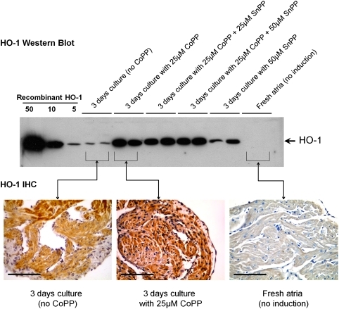
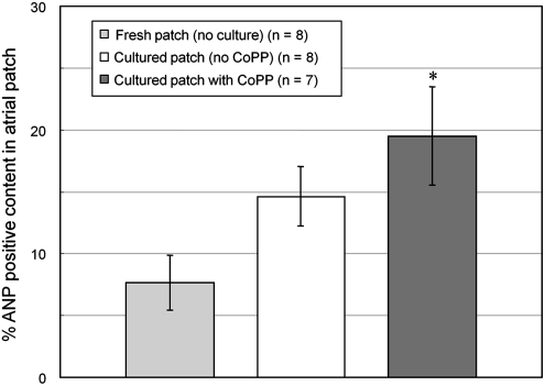
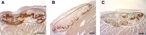
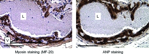
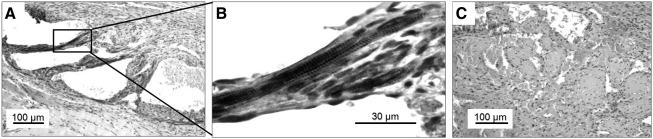
Autologous adult cardiomyocytes are not utilized for heart repair strategies because of their rapid apoptosis after implantation. We examined whether induction of heme oxygenase-1 (HO-1), a mediator of preconditioning, could enhance early postimplant myocyte survival. Three-dimensional 5×5 mm patches of full-thickness adult murine atrial wall, including cardiomyocytes, capillary networks, and extracellular matrix, were cultured with or without HO-1 inducer cobalt protoporphyrin (CoPP), or the HO-1 inhibitor, tin protoporphyrin (SnPP), or both. Patches were then implanted subcutaneously. Freshly procured atrial wall patches implanted without preculturing served as additional controls. By 14 days postimplant, graft cardiomyocyte content was significantly greater in CoPP-treated patches than in either control group (p<0.02). Adult cardiomyocytes did not contract in culture or immediately after implantation. However, by 14 days postimplant, spontaneous contraction had recovered in 47% of CoPP-treated patches, but in only 6% of precultured patches without CoPP, 0% of SnPP-treated patches, and 0% of uncultured patches (p<0.03). CoPP-treated adult cardiomyocyte patches were also observed to remodel spontaneously into endothelial-lined chambers that pumped nonclotting blood. These findings demonstrate that adult cardiomyocytes have more plasticity and capacity for functional recovery than previously recognized and could have application as an autologous cardiomyocyte source for tissue engineering.
Figures
References
-
- Reinecke H. Zhang M. Bartosek T. Murry C.E. Survival, integration, and differentiation of cardiomyocyte grafts: a study in normal and injured rat hearts. Circulation. 1999;100:193. - PubMed
-
- Murry C.E. Jennings R.B. Reimer K.A. Preconditioning with ischemia: a delay of lethal cell injury in ischemic myocardium. Circulation. 1986;74:1124. - PubMed
-
- Bolli R. The late phase of preconditioning. Circ Res. 2000;87:972. - PubMed
-
- Dawn B. Bolli R. HO-1 induction by HIF-1: a new mechanism for delayed cardioprotection? Am J Physiol Heart Circ Physiol. 2005;289:H522. - PubMed
-
- Wu L. Wang R. Carbon monoxide: endogenous production, physiological functions, and pharmacological applications. Pharmacol Rev. 2005;57:585. - PubMed
Publication types
MeSH terms
Substances
Grants and funding
LinkOut - more resources
Full Text Sources
