Review
. 2011 Mar 10;3(1):e00052.
doi: 10.1042/AN20110001.

The curious case of NG2 cells: transient trend or game changer?

Affiliations
Review

The curious case of NG2 cells: transient trend or game changer?

Jean-Marie Mangin et al. ASN Neuro. .

Abstract

It has been 10 years since the seminal work of Dwight Bergles and collaborators demonstrated that NG2 (nerve/glial antigen 2)-expressing oligodendrocyte progenitor cells (NG2 cells) receive functional glutamatergic synapses from neurons (Bergles et al., 2000), contradicting the old dogma that only neurons possess the complex and specialized molecular machinery necessary to receive synapses. While this surprising discovery may have been initially shunned as a novelty item of undefined functional significance, the study of neuron-to-NG2 cell neurotransmission has since become a very active and exciting field of research. Many laboratories have now confirmed and extended the initial discovery, showing for example that NG2 cells can also receive inhibitory GABAergic synapses (Lin and Bergles, 2004) or that neuron-to-NG2 cell synaptic transmission is a rather ubiquitous phenomenon that has been observed in all brain areas explored so far, including white matter tracts (Kukley et al., 2007; Ziskin et al., 2007; Etxeberria et al., 2010). Thus, while still being in its infancy, this field of research has already brought many surprising and interesting discoveries, and has become part of a continuously growing effort in neuroscience to re-evaluate the long underestimated role of glial cells in brain function (Barres, 2008). However, this area of research is now reaching an important milestone and its long-term significance will be defined by its ability to uncover the still elusive function of NG2 cells and their synapses in the brain, rather than by its sensational but transient successes at upsetting the old order established by neuronal physiology. To participate in the effort to facilitate such a transition, here we propose a critical review of the latest findings in the field of NG2 cell physiology--discussing how they inform us on the possible function(s) of NG2 cells in the brain--and we present some personal views on new directions the field could benefit from in order to achieve lasting significance.


Figures
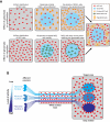
Figure 1
Figure 1. Hypothetical consequences of localized and synchronized release of glutamate on NG2 cells
(A) Cartoon illustrating how synchronized release of glutamate from a localized group of axons or synapses (blue) would affect the behaviour of uniformly distributed NG2 cells (red), taking into account the known effect of glutamate on NG2 cell proliferation (upper three panels), migration/mobility (lower three panel) and differentiation (rightmost panel) (Gallo et al., 1996; Yuan et al., 1998; Gudz et al., 2006). Note that the effects of glutamate on NG2 cell proliferation and migration/mobility would synergize to lower the density of NG2 cells in the area where glutamate is synchronously released. Ultimately, this area will display lower myelination (‘grey matter area’), while the surrounding tissue will exhibit a higher density of NG2 cells and oligodendrocytes (‘white matter area’). (B) Cartoon illustrating how NG2 cells (in red) would behave in different settings where three distinct neuronal nuclei (deep blue to light blue) exhibit a high degree of internal synchronization, but different level of overall activity. Each nucleus projects towards an adjacent target area via a shared white matter tract. In the white matter tract, NG2 cells would tend to accumulate at the boundaries between the bundles of axons corresponding to each nucleus in an activity-dependent manner. In the target grey matter area, an equivalent phenomenon would occur, where NG2 would be depleted from each target area and accumulate at their boundaries in an activity-dependent manner.
Figure 2
Figure 2. Distribution of NG2 cells in the amygdala and corpus callosum of a CNP-GFP (C-type natriuretic peptide-green fluorescent protein) mouse at 7 days after birth (P7)
(AC) High resolution images taken with a standard fluorescence microscope showing the distribution of the proteoglycan NG2 (greyscale in A and B, red in C) and the fluorescent protein EGFP (enhanced green fluorescent protein) expressed under the promoter of the OL-specific protein CNPase (green in C) in a P7 mouse (coronal section). The image in (B) is identical with (A), except for the inclusion of an overlay (in yellow) showing the putative location of several amygdala nuclei, where NG2 is expressed at different levels. The higher magnification image in (C) shows that NG2+CNP-EGFP+ cells are particularly dense in the external and amygdala capsule and that their density varies for each nucleus. Astr: amygdalostriatal transition; BLA: basolateral amygdala nuclei, anterior; BLP: basolateral amygdala nuclei, posterior; CeC: central amygdala nucleus, capsular; CeL: central amygdala nucleus, lateral division; DeN: dorsal endopiriform nucleus; LaDL: lateral amygdala nuclei, dorsolateral; LaVL: lateral amygdala nuclei, ventrolateral; LaVM: lateral amygdala nuclei, ventromedian. (D, E) Single confocal image showing the distribution of NG2+/CNP-EGFP+ cells in corpus callosum (coronal section) of a P7 mouse. The image in (E) corresponds to the area surrounded by the dashed box in (D). Dashed lines in (E) indicate chains of NG2+CNP-EGFP+ cells subdividing adjacent bundles of callosal axons
Figure 3
Figure 3. NG2 cells delimit myelinated bundle of axons in the corpus callosum of a P13 CNP-GFP mouse
(AD) Projection of a small stack of confocal images (five consecutives planes with z = 2 μm) illustrating the tendency of bundles of axons to myelinate [MBP (maltose-binding protein) staining in green in (A), grey in (C)] as a whole independently of surrounding axons. Boundaries are delimited by NG2 cells [NG2 staining in red in (A), grey in (B)] (coronal section). Images in (BD) correspond to the area surrounded by the dashed box in (A). The dashed lines in yellow (BD) indicate the interface between the myelinated bundle of axons labelled for MBP (C) and NG2 cells labelled for the proteoglycan NG2 (B). The image in (D) shows that bright CNP-GFP+ oligodendrocyte processes appear restricted to a single bundle. The yellow asterisk indicates the position of the cell body of one of the oligodendrocyte myelinating the axon bundle delimited by the yellow dashed lines. At this developmental stage, CNP-GFP fluorescence is difficult to detect in NG2 cells, as it is much lower than the fluorescence detected in mature oligodendrocytes.
Figure 4
Figure 4. Hypothetical scenarios of myelination processes occurring either independently between functionally distinct axon bundles or in a random fashion
In this cartoon, two groups of cortical neurons (blue and purple circles) belonging to adjacent cortical columns processing distinct types of sensory information (e.g. somatosensory information from two adjacent digits) project towards homotypic areas of the contralateral cortex via the corpus callosum. In this example, we assume that: (i) callosal axons originating from each group remain segregated (blue and purple lines); (ii) they are delimited by chains of NG2 cells (in red); and (iii) the extent of myelination by individual oligodendrocytes (in green) is limited to individual bundles by NG2 cells. However, other mechanisms could allow similar outcomes. For example, even in the absence of axon segregation and NG2 boundaries, NG2 cells differentiating into oligodendrocytes could initially identify axons belonging to the same functional group via neuron-NG2 cells synapses based on the fact these axons fire synchronously more often than axons carrying distinct types of sensory information. Whatever the exact mechanism, our central argument here is that specifying the myelination process between functionally distinct groups of axons allows to continuously preserve the temporal relationship between action potentials within each group. t1 and t2 indicate the time at which neurons belonging to each group are firing. The firing is strongly synchronized within each group by the fact that they are activated by the same sensory stimulus (e.g. one digit is stimulated at t1, the other at t2). First column from the left: before myelination occurs, assuming that the axonal length is similar for each group of axons (another argument in favour of their bundling) and that their axon diameter is identical, action potentials generated simultaneously in a given group of neurons (blue and purple bars of the left of each schema) should slowly reach their contralateral target at the same time, and their coherence is preserved. Second column from the left: once myelination starts, the conduction velocity of the axons is increased by myelination. If each oligodendrocyte cell limits its myelinating activity to a single bundle (top panel) and tends therefore to myelinate all the axons belonging to the same group at once, the conduction velocity of all axons belonging to this group will be increased in an identical manner, and the synchrony of their action potential will be preserved when they reach their target. On the other hand, if oligodendrocytes myelinate axons they myelinate randomly and as long as myelination is not complete (see third column from the left), each axon would see its conduction velocity increase by a different value and their action potential will be desynchronized once they reach their target.

References

    1. Aguirre A, Chittajallu R, Belachew S, Gallo V. NG2-expressing cells in the subventricular zone are type C-like cells and contribute to interneuron generation in the postnatal hippocampus. J Cell Biol. 2004;165:575–589. - PMC - PubMed
    1. Aguirre A, Dupree JL, Mangin JM, Gallo V. A functional role for EGFR signaling in myelination and remyelination. Nature Neurosci. 2007;10:990–1002. - PubMed
    1. Bakiri Y, Attwell D, Káradóttir R. Electrical signalling properties of oligodendrocyte precursor cells. Neuron Glia Biol. 2009;5:3–11. - PubMed
    1. Banerjee S, Bhat MA. Glial ensheathment of peripheral axons in Drosophila. J Neurosci Res. 2008;86:1189–1198. - PMC - PubMed
    1. Barres BA. The mystery and magic of glia: a perspective on their roles in health and disease. Neuron. 2008;60:430–440. - PubMed