Review
. 2011 Apr;21(2):257-64.
doi: 10.1016/j.sbi.2011.01.003. Epub 2011 Feb 1.

Structural regulation of cullin-RING ubiquitin ligase complexes

Affiliations
Review

Structural regulation of cullin-RING ubiquitin ligase complexes

David M Duda et al. Curr Opin Struct Biol. 2011 Apr.

Abstract

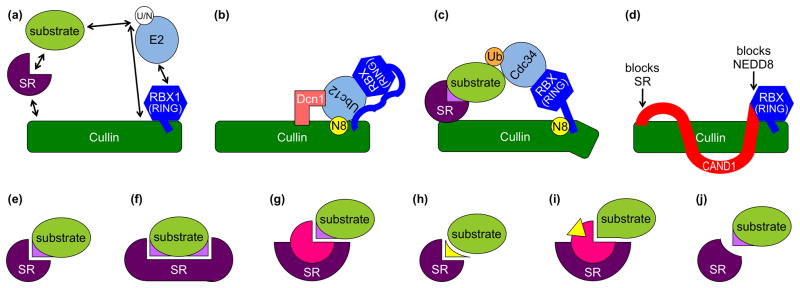
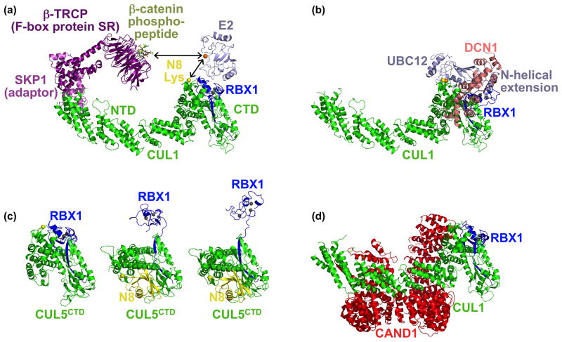
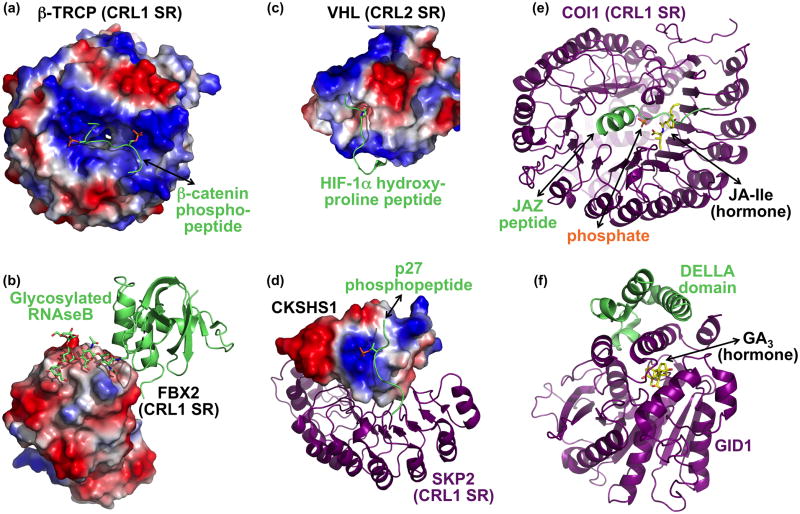
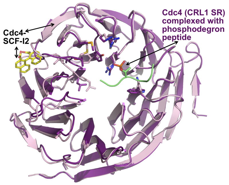
Cullin-RING ligases (CRLs) compose the largest class of E3 ubiquitin ligases. CRLs are modular, multisubunit enzymes, comprising interchangeable substrate receptors dedicated to particular Cullin-RING catalytic cores. Recent structural studies have revealed numerous ways in which CRL E3 ligase activities are controlled, including multimodal E3 ligase activation by covalent attachment of the ubiquitin-like protein NEDD8, inhibition of CRL assembly/activity by CAND1, and several mechanisms of regulated substrate recruitment. These features highlight the potential for CRL activities to be tuned in responses to diverse cellular cues, and for modulating CRL functions through small-molecule agonists or antagonists. As the second installment of a two-review series, this article focuses on recent structural studies advancing our knowledge of how CRL activities are regulated.


Conflict of interest statement

Conflict of interest

The authors declare no competing interest.

Figures

Figure 1
Figure 1. CRL regulation
The elongated cullin scaffold protein (green) interacts via its N-terminal domain (NTD) with a substrate receptor (SR, purple) that recruits substrate (lime). The cullin C-terminal domain (CTD) tightly associates with an RBX1/2 RING protein (blue) that recruits Ubiquitin (Ub) or NEDD8 (N8)-charged E2 enzymes (sky). (a) Conformations and interactions that are regulated are indicated with arrows. (b) Model for dual E3 mechanism for NEDD8 (yellow) ligation to a cullin, which involves both RBX1 and Dcn1 (salmon) cofunctioning as E3s. (c) Model for NEDD8-activated CRL-mediated ubiquitin (orange) ligation to an SR-associated substrate. (d) CAND1 (red) inhibition of cullin-RBX assembly with SRs, and of NEDD8 ligation. (e–j) Regulation of SR-substrate interactions include (e) SR recognition of a specific substrate post-translational modification (violet), (f) SR recognition of multiple specific post-translational modifications (violet) on a substrate, (g) SR binding a partner protein (pink) to recognize a specific substrate post-translational modification (violet), (h) SR binding a small molecule “glue” (yellow) that mediates interactions between the SR and substrate, (i) SR binding a partner protein (pink) that is allosterically modulated by a small molecule hormone (yellow) to then bind a substrate, and (j) inhibition of SR binding after specific post-translational modification (violet) of a substrate.
Figure 2
Figure 2. Conformational control of CRL E3 ligase activities
(a) Structural model of a representative CRL1 complex, obtained by superimposing the Skp1 (violet)-β-TRCP (purple)-β-catenin phosphodegron peptide (lime) and SKP1-F-box domain-CUL1 (green)-RBX1 (blue) structures [5,10]. An E2 (sky) is modeled by superimposing RBX1 RING domain with the UbcH7-bound c-Cbl RING domain [8]. Zinc atoms are shown as grey spheres. Arrows indicate that conformational changes allow the E2 active site cysteine (orange sphere) to approach the substrate peptide or a cullin’s NEDD8 acceptor lysine (yellow sphere). (b) Structural model for the dual E3 mechanism for NEDD8 ligation to cullins [20]. After docking the structure of full-length yeast Ubc12 [20] on the E2-RBX1 model in (a), the RING domain was rotated so that the Ubc12 active site and NEDD8 ligation site are juxtaposed. The second E3 for NEDD8, Dcn1 (salmon), was docked on one side with CUL1 based on a complex structure, and on the other with Ubc12’s N-terminal helical extension based on mutational data [20]. (c) Structures of CUL5CTD-RBX1 and NEDD8~ CUL5CTD-RBX1 showing subdomain rearrangement, including “freeing” of the RBX1 RING domain, in the NEDD8 ligated CRL structures [12]. (d) Structural basis for CAND1-mediated inhibition of CUL1-RBX1. CAND1 (red) locks CRLs in a rigid state, and prevents assembly with SRs and NEDD8 ligation [30].
Figure 3
Figure 3. Selected examples of regulated substrate binding to CRLs
(a) Two precisely positioned basic patches and a peptide binding groove in the CRL1 SR β-TRCP (surface showing electrostatic potential) recruit a doubly-phosphorylated β-catenin (lime, with phosphates as orange/red sticks) [10]. (b) Sugar recognition on a glycosylated substrate (lime) by the CRL1 SR FBX2 (electrostatic surface) [35]. The sugar moiety is depicted in sticks. (c) Recognition of HIF-1α hydroxyproline (peptide in lime, with hydroxyproline in sticks) by the CRL2 SR VHL (electrostatic surface) [32,33]. Only a portion of the VHL structure is shown to highlight substrate recognition. (d) The CRL1 SR Skp2 (purple) imports phosphopeptide recognition by binding to CKSHS1 (electrostatic surface), whose anion binding pocket recognizes the phosphate moiety on the substrate phospho-p27 (lime, with phosphate in orange/red sticks) [39]. (e) The plant hormone jasmonic acid-isoleucine (JA-Ile) functions in a multicomponent molecular glue mechanism to recruit substrates to the CRL1 SR COI-1 (purple). JA-Ile (yellow) mediates binding to the substrate JAZ degron peptide (lime), but the JAZ degron helix also clamps JA-Ile into the COI-1 binding site. A nearby phosphate most likely resembles part of inositol pentakisphosphate, which potentiates interaction between COI-1, JA-Ile, and a JAZ substrate. (f) Gibberellic acid (GA, yellow) uses an indirect allosteric mechanism to promote interactions between DELLA transcriptional repressors (lime) and a multiprotein CRL1 SR that includes GID1 (purple) [42]. GA serves as part of a hydrophobic core required for folding of the GID1 N-terminus. This portion of GID1, in turn, helps recruit DELLA substrates for ubiquitination.
Figure 4
Figure 4. SCF-I2 allosterically inhibits the Cdc4 WD40 β-propeller from binding to a phosphodegron
Superposition of the CRL1 SR Cdc4 WD40 β-propeller from structures of complexes with a phosphodegron peptide (Cdc4 – purple; phosphodegron lime) or with SCF-I2 (Cdc4 – pink; SCF-I2 – yellow). SCF-I2 (yellow) intercalates between two blades of the propeller (pink) that leads to a continuum of structural distortions over 25 Å, ultimately preventing interaction with a phosphodegron.

References

    1. Deshaies RJ, Joazeiro CA. RING domain E3 ubiquitin ligases. Annu Rev Biochem. 2009;78:399–434. - PubMed
    1. Kee Y, Huibregtse JM. Regulation of catalytic activities of HECT ubiquitin ligases. Biochem Biophys Res Commun. 2007;354:329–333. - PMC - PubMed
    1. Petroski MD, Deshaies RJ. Function and regulation of cullin-RING ubiquitin ligases. Nat Rev Mol Cell Biol. 2005;6:9–20. - PubMed
    1. Zimmerman ES, Schulman BA, Zheng N. Structural assembly of cullin-RING ubiquitin ligase complexes. Curr Opin Struct Biol. 2010;20:714–721. - PMC - PubMed
    1. Zheng N, Schulman BA, Song L, Miller JJ, Jeffrey PD, Wang P, Chu C, Koepp DM, Elledge SJ, Pagano M, et al. Structure of the Cul1-Rbx1-Skp1-F boxSkp2 SCF ubiquitin ligase complex. Nature. 2002;416:703–709. - PubMed

Publication types

MeSH terms