. 2011 Jan 15;203(2):258-62.
doi: 10.1093/infdis/jiq028. Epub 2010 Dec 9.

Lack of detectable HIV-1-specific CD8(+) T cell responses in Zambian HIV-1-exposed seronegative partners of HIV-1-positive individuals

Affiliations

Lack of detectable HIV-1-specific CD8(+) T cell responses in Zambian HIV-1-exposed seronegative partners of HIV-1-positive individuals

Marylyn M Addo et al. J Infect Dis. .

Abstract

Human immunodeficiency virus type 1 (HIV-1)-specific T cell responses were characterized in a blinded study involving infected individuals and their seronegative exposed uninfected (EU) partners from Lusaka, Zambia. HIV-1-specific T cell responses were detected ex vivo in all infected individuals and amplified, on average, 27-fold following in vitro expansion. In contrast, no HIV-1-specific T cell responses were detected in any of the EU partners ex vivo or following in vitro expansion. These data demonstrate that the detection of HIV-1-specific T cell immunity in EU individuals is not universal and that alternative mechanisms may account for protection in these individuals.


Figures
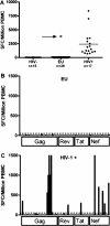
Figure 1.
Figure 1.
Human immunodeficiency virus type 1 (HIV-1)–specific T cell responses were measured in blinded fashion by ex vivo interferon–γ enzyme-linked immunosorbent spot (ELISPOT) assay in HIV-1 seropositive and exposed seronegative individuals. ELISPOT data were available for all 61 study subjects. Panel A shows strong T cell responses to HIV-1 clade C Gag, Rev, Tat, and Nef overlapping peptides in all individuals identified as known HIV-seropositive study participants after unblinding. Results are shown as total magnitude of SFC/106 peripheral blood mononuclear cells (PBMCs). In contrast, study subjects who were identified as HIV-1 seronegative at study entry (including 28 high-risk exposed uninfected [EU] and 15 low-risk HIV seronegative control subjects) did not have detectable HIV-1 clade C–specific T cell responses ex vivo. One individual (AD45; arrow) who was classified as EU at study entry by an enzyme-linked immunosorbent assay (ELISA) result that was negative for HIV at 1 month prior to phlebotomy had detectable T cell responses by ex vivo ELISPOT. This individual had a positive HIV ELISA result at 7 weeks after phlebotomy and was sampled and assayed during acute HIV infection (confirmed by isolation of replication-competent virus from a PBMC sample from same time point as the ELISPOT assay; no plasma was available for HIV viral load quantification). Panels B and C show representative examples of ELISPOT screenings with overlapping peptides spanning HIV-1 clade C Gag, Rev, Tat, and Nef in an HIV-1–seropositive individual and a high-risk exposed seronegative individual, respectively. All experiments were performed blinded. Positive responses were defined as at least 3 times the number of SFCs in the control wells and had to be >50 SFC/106 PBMCs [10].
Figure 2.
Figure 2.
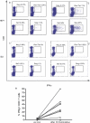
Large expansion of human immunodeficiency virus type 1 (HIV-1)–specific CD8+ T cell responses measured by flow cytometry in HIV-1–positive (but not HIV-1 high- or low-risk seronegative) individuals after HIV-1–specific in vitro stimulation and expansion. HIV-1 clade C Gag, Rev, Tat, and Nef responses were measured by flow cytometry ex vivo and after 10-day in vitro peptide-specific stimulation with pools of overlapping peptides in a subset of HIV-positive (n = 8) and HIV-seronegative individuals (n = 8), including 4 high-risk and 4 low-risk EU. Representative examples of the total HIV-1–specific intracellular interferon–γ response ex vivo (A and C) and after 10-day stimulation (B and D) for an HIV-1–positive individual (A and B) and an exposed HIV-1–uninfected individual (EU) (C and D) are shown. Panel E shows the summary for the total HIV-1–specific T cell responses for 16 individuals tested pre- and post-stimulation as measured by flow cytometry. HIV-1–specific CD8+ T cell responses were detectable in all HIV-1–infected individuals directly ex vivo, with the total response ranging from 0.34% to 4.06% (median response, 1%). These virus-specific CD8+ T cell responses expanded significantly after in vitro stimulation (median increase, 27-fold; range, 9–69-fold; P = .002), reaching median frequencies of total HIV-1–specific CD8+ T cells of 33% (range, 6%–80%). No HIV-1–specific T cell responses above background were detected by intracellular cytokine staining in the 8 HIV-1–negative individuals (4 low-risk and 4 high-risk seronegative subjects) who were tested directly ex vivo or following the in vitro expansion (C, D, and E), as represented by the graph line overlying the x-axis, showing no change between before and after stimulation (E).

References

    1. Miyazawa M, Lopalco L, Mazzotta F, Lo Caputo S, Veas F, Clerici M. The ‘immunologic advantage’ of HIV-exposed seronegative individuals. AIDS. 2009;23:161–175. - PubMed
    1. Alimonti JB, Kimani J, Matu L, et al. Characterization of CD8 T-cell responses in HIV-1-exposed seronegative commercial sex workers from Nairobi, Kenya. Immunol Cell Biol. 2006;84:482–485. - PubMed
    1. Makedonas G, Bruneau J, Lin H, Sekaly RP, Lamothe F, Bernard NF. HIV-specific CD8 T-cell activity in uninfected injection drug users is associated with maintenance of seronegativity. AIDS. 2002;16:1595–1602. - PubMed
    1. Kaul R, Rowland-Jones SL, Kimani J, et al. Late seroconversion in HIV-resistant Nairobi prostitutes despite pre-existing HIV-specific CD8+ responses. J Clin Invest. 2001;107:341–349. - PMC - PubMed
    1. Hladik F, Desbien A, Lang J, et al. Most highly exposed seronegative men lack HIV-1–specific, IFN-gamma-secreting T cells. J Immunol. 2003;171:2671–2683. - PubMed

Publication types