. 2011 Mar 25;286(12):10066-72.
doi: 10.1074/jbc.M110.185082. Epub 2011 Feb 2.

Ectodomain shedding and autocleavage of the cardiac membrane protease corin

Affiliations

Ectodomain shedding and autocleavage of the cardiac membrane protease corin

Jingjing Jiang et al. J Biol Chem. .

Abstract

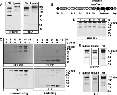
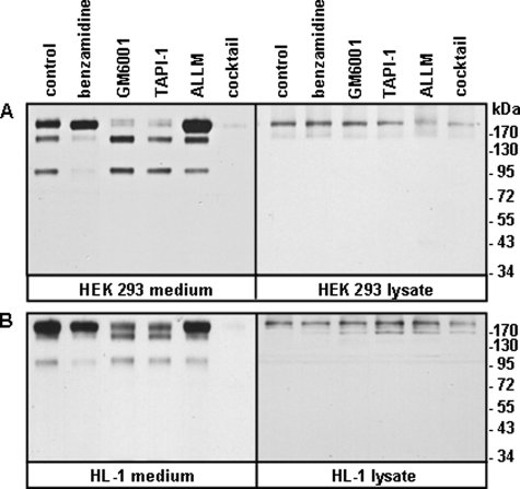
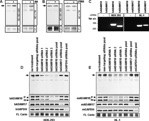
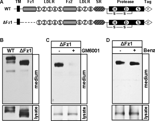
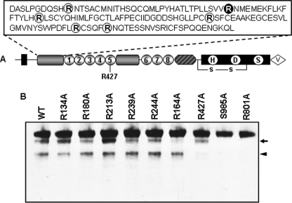
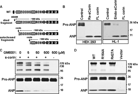
Corin is a cardiac membrane protease that activates natriuretic peptides. It is unknown how corin function is regulated. Recently, soluble corin was detected in human plasma, suggesting that corin may be shed from cardiomyocytes. Here we examined soluble corin production and activity and determined the proteolytic enzymes responsible for corin cleavage. We expressed human corin in HEK 293 cells and detected three soluble fragments of ∼180, ∼160, and ∼100 kDa, respectively, in the cultured medium by Western blot analysis. All three fragments were derived from activated corin molecules. Similar results were obtained in HL-1 cardiomyocytes. Using protease inhibitors, ionomycin and phorbol myristate acetate stimulation, small interfering RNA knockdown, and site-directed mutagenesis, we found that ADAM10 was primarily responsible for shedding corin in its juxtamembrane region to release the ∼180-kDa fragment, corresponding to the near-entire extracellular region. In contrast, the ∼160- and ∼100-kDa fragments were from corin autocleavage at Arg-164 in frizzled 1 domain and Arg-427 in LDL receptor 5 domain, respectively. In functional studies, the ∼180-kDa fragment activated atrial natriuretic peptide, whereas the ∼160- and ∼100-kDa fragments did not. Our data indicate that ADAM-mediated shedding and corin autocleavage are important mechanisms regulating corin function and preventing excessive, potentially hazardous, proteolytic activities in the heart.


Figures

FIGURE 1.
FIGURE 1.
Detection of soluble corin fragments. A, recombinant human corin proteins in the conditioned medium (CM) and cell lysate (Lysate) from transfected HEK 293 and HL-1 cells were analyzed by Western blotting. B, corin protein domains. The transmembrane (TM), frizzled-like (Fz), LDL receptor (LDLR), scavenger receptor (SR), and protease domains are shown. The activation cleavage site, active site residues His (H), Asp (D), and Ser (S), C-terminal V5 tag (V), and the disulfide bond connecting the propeptide and the protease domain are indicated. C, soluble corin fragments in the medium from HEK 293 (top) or HL-1 (bottom) cells expressing recombinant (re) corin collected over time were analyzed by Western blotting under non-reducing and reducing conditions. The ∼40-kDa bands in the right panels represent the activated corin protease domain. D, the conditioned medium was collected from transfected HEK 293 cells and incubated at 37 °C over time. Corin fragments were analyzed by Western blotting. E and F, human, mouse and rat soluble corin fragments in the conditioned medium from transfected HEK 293 and HL-1 cells, respectively. Data are from representative experiments repeated at least three times.
FIGURE 2.
FIGURE 2.
Effects of protease inhibitors. HEK 293 (A) and HL-1 (B) cells expressing recombinant human corin were incubated with vehicle (control), benzamidine, GM6001, TAPI-1, N-acetyl-leucyl-leucyl-methionine, or protease inhibitor mixture. Soluble corin fragments in the conditioned medium were analyzed by Western blotting (left panels). Corin proteins in cell lysate were verified (right panels). Data are from representative experiments repeated at least three times.
FIGURE 3.
FIGURE 3.
ADAM-mediated corin shedding. HEK 293 and HL-1 cells expressing recombinant corin were treated with vehicle, ionomycin (IM) (A) or PMA (B). Corin fragments in the conditioned medium (CM) and cell lysate (LS) were analyzed by Western blotting. C, expression of human (h) and mouse (m) ADAM10 and ADAM17 mRNAs in parental HEK 293 and HL-1 cells was confirmed by RT-PCR. HEK 293 (D) and HL-1 (E) cells expressing corin were transfected with single or pooled siRNAs against the human or mouse ADAM10 or ADAM17 genes. The production of the ∼180-kDa fragment (arrows) in the medium was reduced in ADAM10 siRNA-treated cells, as shown by Western blotting. Controls included non-targeting siRNAs pool or siRNAs pools against the human and mouse GAPDH genes. The reduction of ADAM10 (proform (P) and mature form (m) (arrow heads)), ADAM17, and GAPDH proteins in transfected cells was shown by Western blotting. Full-length (FL) corin in cell lysate also was shown. Data are from representative experiments that were repeated at least three times.
FIGURE 4.
FIGURE 4.
ADAM-mediated cleavage in the juxtamembrane domain. A, illustration of WT corin and the Fz1 deletion mutant (ΔFz1). HEK 293 cells expressing WT corin and the ΔFz1 mutant incubated without (B) or with GM6001 (C) or benzamidine (Benz) (D). Soluble corin fragments in the medium were analyzed by Western blotting. Lysate was included as a control. Data are from representative experiments that were repeated at least three times.
FIGURE 5.
FIGURE 5.
Soluble corin fragments from the S985A and R801A mutants. A, illustration of WT corin, active site mutant S985A, and activation cleavage site mutant R801A. Soluble corin fragments from transfected HEK 293 (B) and HL-1 (C) cells expressing WT corin and the S985A and R801A mutants were analyzed by Western blotting (left panels). Cell lysate was included as a control for corin expression levels (right panels). Data are from representative experiments that were repeated at least three times.
FIGURE 6.
FIGURE 6.
Corin autocleavage sites. A, illustration of Arg residues in the Fz1 and LDLR5 domains that were substituted by Ala in single mutants. B, soluble corin fragments in the medium from HEK 293 cells expressing WT corin and mutants were analyzed by Western blotting. The S985A and R801A mutants, in which autocleavage was abolished, were used as controls. The ∼160- and ∼100-kDa bands are indicated by an arrow or arrow head, respectively. Data are from representative experiments that were repeated at least three times.
FIGURE 7.
FIGURE 7.
The activity of soluble corin fragments. A, illustration of soluble corin fragments. B, pro-ANP processing activity. Conditioned medium containing recombinant (Re) or endogenous (Endo) soluble (s) corin from HEK 293 (left) or HL-1 (right) cells were incubated with pro-ANP. Control medium and cell lysate with full-length (FL) corin were included as negative and positive controls, respectively. Pro-ANP to ANP conversion was analyzed by Western blotting. C, HEK 293 cells expressing corin or parental HEK 293 cells were incubated with increasing doses of GM6001. Soluble corin fragments (top) and pro-ANP processing activity (bottom) in the medium were analyzed by Western blotting. D, soluble corin fragments (top) and pro-ANP processing activity (bottom) in the conditioned medium from HEK 293 cells expressing WT corin and mutants R164A and R427A were analyzed by Western blotting. Data are from representative experiments that were repeated at least three times.

References

    1. McGrath M. F., de Bold M. L., de Bold A. J. (2005) Trends Endocrinol. Metab. 16, 469–477 - PubMed
    1. Potter L. R., Abbey-Hosch S., Dickey D. M. (2006) Endocr. Rev. 27, 47–72 - PubMed
    1. Yan W., Sheng N., Seto M., Morser J., Wu Q. (1999) J. Biol. Chem. 274, 14926–14935 - PubMed
    1. Yan W., Wu F., Morser J., Wu Q. (2000) Proc. Natl. Acad. Sci. U.S.A. 97, 8525–8529 - PMC - PubMed
    1. Chan J. C., Knudson O., Wu F., Morser J., Dole W. P., Wu Q. (2005) Proc. Natl. Acad. Sci. U.S.A. 102, 785–790 - PMC - PubMed

Publication types

MeSH terms