. 2011 Apr;107(5):843-53.
doi: 10.1093/aob/mcr005. Epub 2011 Feb 2.

Three-dimensional distribution of vessels, passage cells and lateral roots along the root axis of winter wheat (Triticum aestivum)

Affiliations

Three-dimensional distribution of vessels, passage cells and lateral roots along the root axis of winter wheat (Triticum aestivum)

Haiwen Wu et al. Ann Bot. 2011 Apr.

Abstract

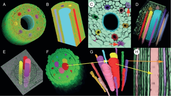
Background and aims: The capacity of a plant to absorb and transport water and nutrients depends on anatomical structures within the roots and their co-ordination. However, most descriptions of root anatomical structure are limited to 2-D cross-sections, providing little information on 3-D spatial relationships and hardly anything on their temporal evolution. Three-dimensional reconstruction and visualization of root anatomical structures can illustrate spatial co-ordination among cells and tissues and provide new insights and understanding of the interrelation between structure and function.

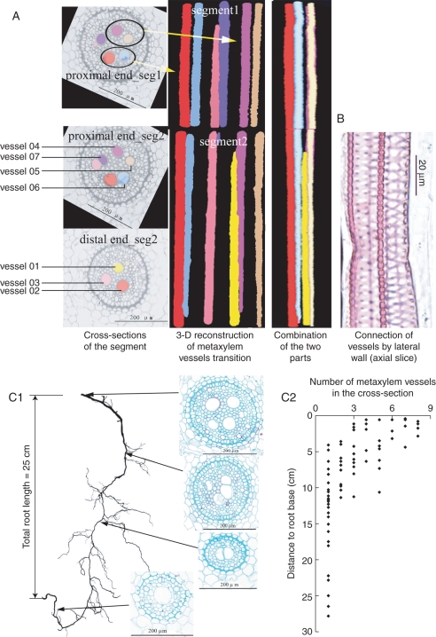
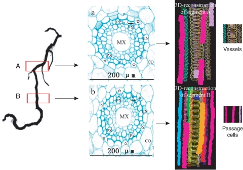
Methods: Classical paraffin serial-section methods, image processing, computer-aided 3-D reconstruction and 3-D visualization techniques were combined to analyse spatial relationships among metaxylem vessels, passage cells and lateral roots in nodal roots of winter wheat (Triticum aestivum).

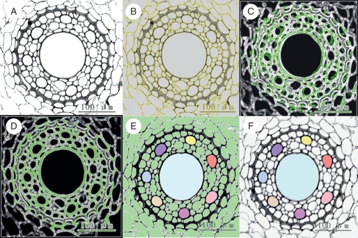
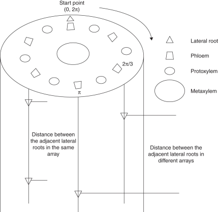
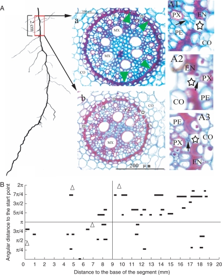
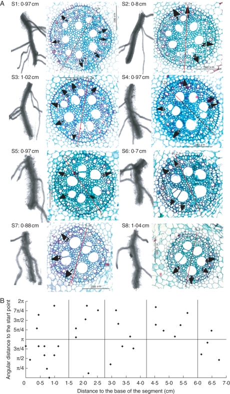
Key results: 3-D reconstruction demonstrated that metaxylem vessels were neither parallel, nor did they run directly along the root axis from the root base to the root tip; rather they underwent substitution and transition. Most vessels were connected to pre-existent or newly formed vessels by pits on their lateral walls. The spatial distributions of both passage cells and lateral roots exhibited similar position-dependent patterns. In the transverse plane, the passage cells occurred opposite the poles of the protoxylem and the lateral roots opposite those of the protophloem. Along the axis of a young root segment, the passage cells were arranged in short and discontinuous longitudinal files, thus as the tissues mature, the sequence in which the passage cells lose their transport function is not basipetal. In older segments, passage cells decreased drastically in number and coexisted with lateral roots. The spatial distribution of lateral roots was similar to that of the passage cells, mirroring their similar functions as lateral pathways for water and nutrient transport to the stele.

Conclusions: With the 3-D reconstruction and visualization techniques developed here, the spatial relationships between vessels, passage cells and lateral roots and the temporal evolution of these relationships can be described. The technique helps to illustrate synchronization and spatial co-ordination among the root's radial and axial pathways for water and nutrient transport and the interdependence of structure and function in the root.


Figures

Fig. 1.
Fig. 1.
Main steps of image processing in the 3-D reconstruction program (xylem vessels as an example). (A) Colour images of the root stele are converted to grey-scale images, then loaded into the AMBIOS program and adjusted to optimal brightness and definition. (B) Edges of cells are computed by the program after setting up the appropriate parameters. The yellow curves are the computed cell edges (coinciding with the cell walls). (C) An image in the middle of the series is defined as the reference image and the cell edges are coloured green. A succeeding or anteceding image (cell edges coloured white) is superimposed. The image does not match the reference image until the green and the white lines coincide. (D) Images are rotated and translated to best match the reference. (E) On the image in the middle of the series, all the cells are filled with green (default colour of the system). Xylem vessels in the reference image to be reconstructed are selected and their lumens are manually filled with different colours. The vessel colour registrations are then propagated to the other aligned images. (F) The background colour, no longer necessary, is removed leaving the selections (vessels) for 3-D-reconstruction and visualization.
Fig. 2.
Fig. 2.
Representations of 3-D distribution of lateral roots. Since lateral roots originate from the pericycle cells out of the phloem, the phloem bundles are considered as the reference for the lateral roots. One phloem bundle is arbitrarily taken as the starting point. The others are labelled clockwise, being identified by their radial angles from the start point. In the longitudinal direction, the position of a lateral root is noted by its distance from the base of the segment. Thus the 3-D position of any lateral root is defined by a radial angle in transverse section and the axial distance to the segment base.
Fig. 3.
Fig. 3.
Various 3-D views of the reconstructed root tissues. The green colour represents living cells in the stele and other colours represent vessels. (A) The 3-D reconstruction of the stele with the centre metaxylem vessel shown as open. The thickened cell walls of the endodermis cells are coloured brown. (B) Two perpendicular cutting profiles on the reconstructed stele, with narrow protoxylem vessels at the periphery and a wide metaxylem vessel in the centre. (C) A 3-D reconstruction of the stele with six protoxylem vessels and one central metaxylem vessel. Note passage cells (red arrows) opposite the protoxylem poles. (D) Compound views of vessels including a cut view (note the metaxylem vessel in blue) and the view of the edges as wireframes. Solid forms inside the wireframes indicate the centre of gravity of the vessels. (E) Reconstructed metaxylem vessels running through a cross-section show their relative position in the stele. (F) Reconstructed stele of a nodal root. Large vessels are metaxylem and narrow ones are protoxylem. Note that there are more than one metaxylem vessel in the stele. (G) Reconstruction and visualization of isolated metaxylem and protoxylem vessels by VRML file based on the same serial cross-sections as in (F). (H) Reconstructed longitudinal profile of the vessels based on the same serial cross-sections as in (F). Arrows with the same colour in (F), (G) and (H) indicate the same vessels.
Fig. 4.
Fig. 4.
3-D visualization underlines the substitution and transitions of metaxylem vessels along the wheat nodal root axis. (A) Cross-sections of the stele and 3-D reconstructions and visualization of the vessels. As the presence of a lateral root interrupts the progressive change of anatomical structure of the main root, the 3-D reconstruction and visualization of the root segment have to be performed separately on two parts: segment 1 towards the root base and segment 2 to the root apex. On the left are presented the cross-sections of two root segments: proximal end (towards the root base) of segment 1, proximal end of segment 2 and distal end (towards the root apex) of segment 2. The images in the middle of (A) are 3-D reconstructions in each segment, illustrating the transition of vessels along the root segment. Vessels in the segments can be distinguished one from the other by colours, note that vessel 07 (violet) that was visible in segment 1 and in the proximal end of segment 2, did not appear in the distal end of segment 2, meanwhile the yellow one (vessel 01) was newly formed, as it does not occur in segment 1. On the right of (A) is a side view of the two segments performed by section interpolation described within a VRML file. (B) Longitudinal section showing two vessels are connected by pits on their lateral walls. (C1) Evolution of the metaxylem vessels along a nodal root axis. The length of the root is 25 cm. The slice images are taken at each segment indicated by the arrows. The number of metaxylem vessels decreases from root base to root tip. (C2) Evolution of the number of metaxylem vessels along the root axis – data from cross-sections of 12 roots. In most roots, the number of metaxylem vessels decreases to one at 10 cm from the root base.
Fig. 5.
Fig. 5.
3-D arrangement of passage cells on root segments close to the root tip. Left: location of the samples. Root segments about 0·5 cm long were taken at 3-cm intervals (segment A, more proximal to the root base) and 1·5 cm (segment B, more distal) to the root tip, the lengths of the reconstructed segments were 500 µm for segment A and 590 μm for segment B. Middle: cross-sections made on segments A and B, respectively. MX, Metaxylem vessel; PX, protoxylem vessel; CO, cortex; EN, endodermis. Six passage cells (indicated by stars and differing from the other endodermal cells by having non-thickened inner-tangential walls) are in (b) and only three in (a). Note that pericycle cells adjacent to passage cells (indicated by the arrows) are smaller than the others. Right: 3-D-reconstructions of segments A and B showing short non-continuous files of passage cells represented by different colour solid columns surrounding the metaxylem vessels (wireframes).
Fig. 6.
Fig. 6.
Distribution of passage cells in an old root segment near the root base. (a) Cross-sections at about 14 cm (a) and 12 cm (b) from the root tip. Note that the passage cells (indicated by stars) are much less frequent in the section taken near the root base (a) than in the section from the more distal position (b). The green arrowheads indicate the radial position of the four lateral roots on the 2-cm-long segment. (A1), (A2) and (A3) are magnifications of portions of selected sections. Several (A1) or one (A2) small pericycle cells (indicated by black arrows) lie between the protoxylem poles and the passage cells. Some of them are very small and look similar to an intercellular space (A3). CO, Cortex; EN, endodermis; PE, pericycle; MX, metaxylem; PX, protoxylem. (B) The longitudinal arrangement of passage cells along the root axis. Passage cells are arranged in short discontinuous files. They tend to cluster in one half of the transverse plane over a short distance (in 0-π for 0–9 mm from the root base; in π-2π for 9–19 mm from the root base). In the longer distance, passage cells seem to disperse radially. Triangles indicate the positions of the lateral roots.
Fig. 7.
Fig. 7.
Spatial arrangements of lateral roots along nodal root axis. (A) Projections of lateral roots on the transverse plane (black triangles). A 7-cm-long root was cut into eight segments labelled s1–s8, with s1 being the most proximal (nearest to the root base), and s8 the most distal (nearest to the root tip). Scan pictures and the corresponding cross-section images of each segment are lined up. The values following the segment codes are their respective lengths. Note that most lateral roots occur on the convex side of the mother root. Numbers following the black triangles indicate the sequence of the lateral roots on the segment in the direction of root base to tip. (B) The radial and axial distribution of lateral roots. Over short distances (within the two vertical lines), lateral roots tend to cluster on one side of a transverse plane, but over longer distances, there is a tendency for a more uniform distribution. See Fig. 2 for the labelling of lateral root position on the mother roots. The red lines in the cross-sections serve as the starting points (0, 2π) and central axes for aligning the serial sections.

References

    1. Aloni R. The control of vascular differentiation. Journal of Plant Science. 1992;153:S90–S92.
    1. Aloni R. The induction of vascular tissue by auxin. In: Davies PJ, editor. Plant hormones: biosynthesis, signal transduction, action. Berlin: Springer; 2004.
    1. Aloni R, Zimmermann MH. The control of vessel size and density along the plant axis: a new hypothesis. Differentiation. 1983;24:203–208.
    1. Aloni R, Aloni E, Langhans M, Ullrich CI. Role of cytokinin and auxin in shaping root architecture: regulating vascular differentiation, lateral root initiation, root apical dominance and root gravitropism. Annals of Botany. 2006;97:883–893. - PMC - PubMed
    1. Bardage LS. Three-dimensional modeling and visualization of whole Norway spruce latewood tracheids. Wood and Fiber Science. 2001;33:627–638.

Publication types