. 2011 Feb 1;25(3):232-7.
doi: 10.1101/gad.2001911.

Tyrosine phosphorylation controls brassinosteroid receptor activation by triggering membrane release of its kinase inhibitor

Affiliations

Tyrosine phosphorylation controls brassinosteroid receptor activation by triggering membrane release of its kinase inhibitor

Yvon Jaillais et al. Genes Dev. .

Abstract

Receptor tyrosine kinases control many critical processes in metazoans, but these enzymes appear to be absent in plants. Recently, two Arabidopsis receptor kinases--BRASSINOSTEROID INSENSITIVE 1 (BRI1) and BRI1-ASSOCIATED KINASE1 (BAK1), the receptor and coreceptor for brassinosteroids--were shown to autophosphorylate on tyrosines. However, the cellular roles for tyrosine phosphorylation in plants remain poorly understood. Here, we report that the BRI1 KINASE INHIBITOR 1 (BKI1) is tyrosine phosphorylated in response to brassinosteroid perception. Phosphorylation occurs within a reiterated [KR][KR] membrane targeting motif, releasing BKI1 into the cytosol and enabling formation of an active signaling complex. Our work reveals that tyrosine phosphorylation is a conserved mechanism controlling protein localization in all higher organisms.


Figures
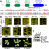
Figure 1.
Figure 1.
[KR][KR] repeats target BKI1 to the PM. (A) Schematic representation of BKI1 conserved regions and alignment between membrane targeting regions of BKI1 orthologs. (Red) Doublets of dibasic residues; (blue) Y211. (B) Deletion constructs used to identify the BKI1 targeting region. (C) Identification of key residues within the membrane targeting motif required for localization. (RKKK) RK179–180 and KK197–198 mutation into AAAA; (KKKR) KK209–210 and KR 218–219 into AAAA. (D) BKI1 gain-of-function phenotype; rosette radius in millimeters ± SD (n = 25). (E) Expression level of transgenic proteins. Bar 10 μm.
Figure 2.
Figure 2.
Tyrosine phosphorylation regulates BKI1 membrane localization in response to BRs. (A,B) Immunoprecipitation (IP) of BKI1-mCITRINE with an anti-GFP antibody and immunoblot (IB) using an anti-GFP antibody or anti-phosphotyrosine antibody (pY). (C) Tyrosine phosphorylation of in vitro translated BKI1-Flag/BKI1Y211F-Flag in the presence of BRI1-KD(814–1196) or BRI1-KD(814–1196) D1027N. (D) Subcellular localization and dynamics upon BR treatment of the Y211F and Y211D mutant proteins. Myri designates protein with a myristoylation signal. (E) Overexpression phenotype of BKI1 tyrosine mutant proteins compared with wild type and myriBKI1; rosette radius in millimeters ± SD (n = 25). (F) Expression level of transgenic proteins. Bar 10 μm.
Figure 3.
Figure 3.
BKI1-CT is required for BRI1 interaction and inhibition, and for BKI1 phosphorylation. (A) Overexpression phenotype of BKI1-CT deletion or point mutant proteins; rosette radius in millimeters ± SD (n = 25). (B) Expression level of transgenic proteins. (C) Coimmunoprecipitation of BKI1 wild-type and mutant versions with BRI1-6xHA. Localization (D) and tyrosine phosphorylation (E) of BKI1-CT mutant proteins compared with wild-type BKI1. Bar 10 μm.
Figure 4.
Figure 4.
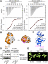
Mechanism of BRI1 inhibition by the BKI1 C-terminal tail. (A) ITC of BRI1-KD(865–1160) versus BKI1-CT, and BRI1-KD(865–1160) D1027N versus MAKR1-CT. Shown are experimental values ± fitting errors. (B) Sequence divergence between BRI1 and BRL1 mapped onto the molecular surface of a BRI1 homology model. Surfaces are colored according to sequence conservation, orange to white for variable to invariant residues. (C) Ribbon diagram of the BRI1 homology model depicting the position of the nucleotide (in bond representations) and the C-lobe mutations (in magenta). (D) Coimmunoprecipitation of BAK1-mCHERRY with BRI1-mCITRINE in the presence of 20 μM BKI1-CT peptide, LQII peptide, or buffer. (E) MAKR1 gain-of-function phenotype; rosette radius in millimeters ± SD (n = 25).

References

    1. Bayliss R, Sardon T, Vernos I, Conti E 2003. Structural basis of Aurora-A activation by TPX2 at the mitotic spindle. Mol Cell 12: 851–862 - PubMed
    1. Belkhadir Y, Chory J 2006. Brassinosteroid signaling: A paradigm for steroid hormone signaling from the cell surface. Science 314: 1410–1411 - PubMed
    1. Cock JM, Vanoosthuyse V, Gaude T 2002. Receptor kinase signalling in plants and animals: Distinct molecular systems with mechanistic similarities. Curr Opin Cell Biol 14: 230–236 - PubMed
    1. Diella F, Haslam N, Chica C, Budd A, Michael S, Brown NP, Trave G, Gibson TJ 2008. Understanding eukaryotic linear motifs and their role in cell signaling and regulation. Front Biosci 13: 6580–6603 - PubMed
    1. Geldner N, Hyman DL, Wang X, Schumacher K, Chory J 2007. Endosomal signaling of plant steroid receptor kinase BRI1. Genes Dev 21: 1598–1602 - PMC - PubMed

Publication types

MeSH terms