. 2011 Apr;22(7):1070-9.
doi: 10.1091/mbc.E10-08-0731. Epub 2011 Feb 2.

Coilin participates in the suppression of RNA polymerase I in response to cisplatin-induced DNA damage

Affiliations

Coilin participates in the suppression of RNA polymerase I in response to cisplatin-induced DNA damage

Andrew S Gilder et al. Mol Biol Cell. 2011 Apr.

Abstract

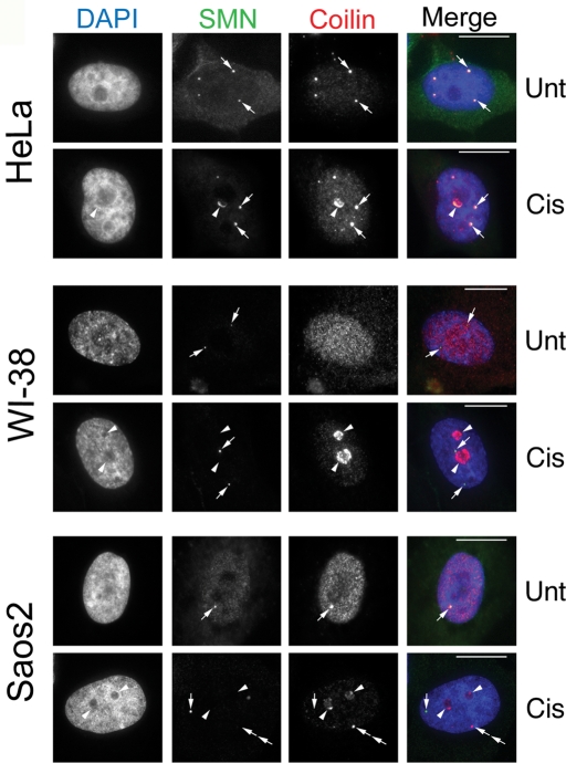
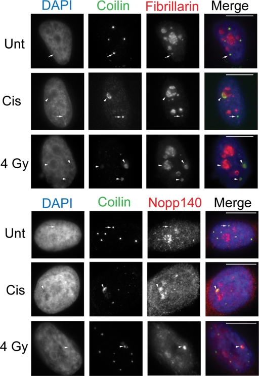
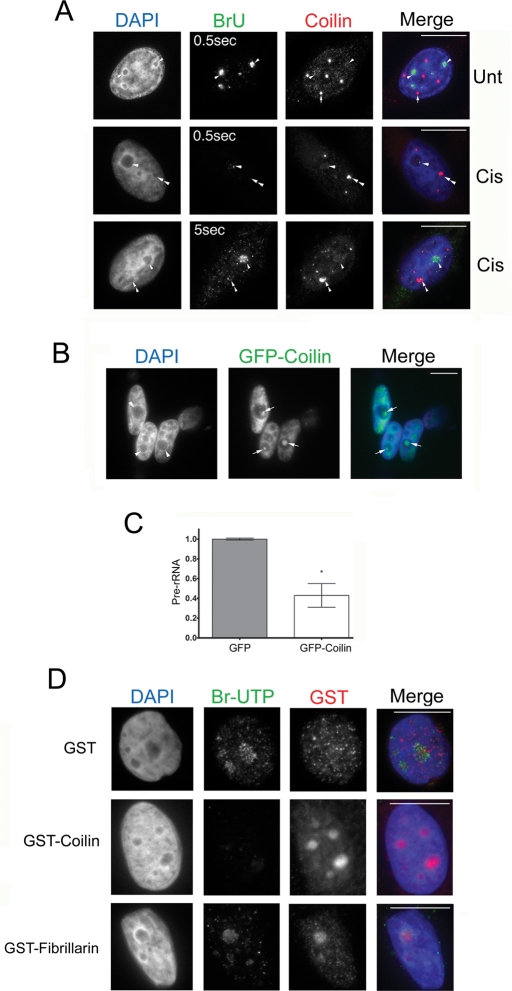
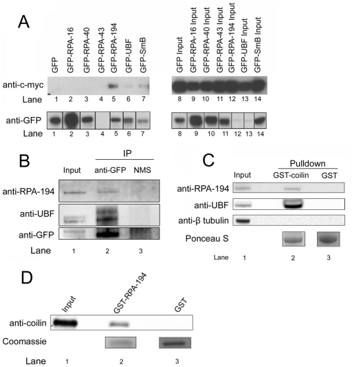
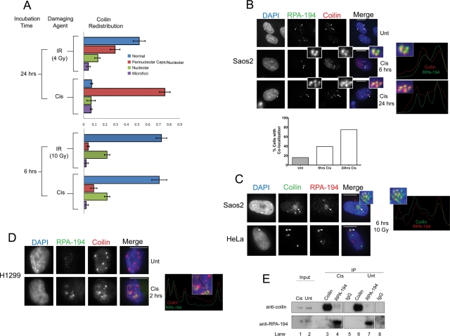
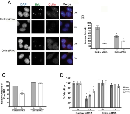
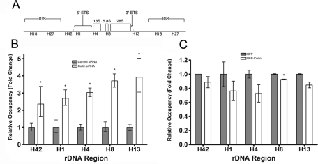
Coilin is a nuclear phosphoprotein that concentrates within Cajal bodies (CBs) and impacts small nuclear ribonucleoprotein (snRNP) biogenesis. Cisplatin and γ-irradiation, which cause distinct types of DNA damage, both trigger the nucleolar accumulation of coilin, and this temporally coincides with the repression of RNA polymerase I (Pol I) activity. Knockdown of endogenous coilin partially overrides the Pol I transcriptional arrest caused by cisplatin, while both ectopically expressed and exogenous coilin accumulate in the nucleolus and suppress rRNA synthesis. In support of this mechanism, we demonstrate that both cisplatin and γ-irradiation induce the colocalization of coilin with RPA-194 (the largest subunit of Pol I), and we further show that coilin can specifically interact with RPA-194 and the key regulator of Pol I activity, upstream binding factor (UBF). Using chromatin immunoprecipitation analysis, we provide evidence that coilin modulates the association of Pol I with ribosomal DNA. Collectively, our data suggest that coilin acts to repress Pol I activity in response to cisplatin-induced DNA damage. Our findings identify a novel and unexpected function for coilin, independent of its role in snRNP biogenesis, establishing a new link between the DNA damage response and the inhibition of rRNA synthesis.


Figures

FIGURE 1:
FIGURE 1:
Cisplatin-induced DNA damage triggers nucleolar coilin accumulation. HeLa, WI-38, and Saos2 cells were untreated (Unt) or treated (Cis) for 24 h with cisplatin and immunostained for coilin (red) and SMN (green). Nuclei were stained by 4′,6-diamino-2-phenylindole (DAPI) (blue). For HeLa cells, arrows denote CBs containing both SMN and coilin. Arrowheads mark nucleolar accumulation of coilin and SMN. For WI-38 cells, SMN foci lacking coilin (Gems) are indicated by arrows, and arrowheads mark nucleolar coilin. For Saos2 cells, arrows in the untreated cell denote a CB. Arrows in the cisplatin-treated cells mark a Gem while arrowheads show nucleolar coilin. Double arrows represent an SMN-negative coilin focus. Scale bars = 10 μm.
FIGURE 2:
FIGURE 2:
Fibrillarin and Nopp140 redistribute with coilin in response to cisplatin and γ-irradiation. HeLa cells were untreated (Unt) or treated with cisplatin or γ-irradiation (4 Gy) and immunostained for coilin, fibrillarin, and Nopp140 after 24 h. DAPI staining was used to detect the nucleus. Arrows mark CBs. Arrowheads mark coilin and fibrillarin or Nopp140 nucleolar redistributions. Scale bars = 10 μm.
FIGURE 3:
FIGURE 3:
Nucleolar coilin suppresses Pol I transcription. (A) Coilin-occupied nucleoli are associated with reduced BrU incorporation. Untreated or cisplatin-treated (24 h) HeLa cells were incubated in 2 mM BrU for 2 h followed by detection of Pol I transcripts with anti-BrdU antibodies (green) and coilin with anti-coilin antibodies (red) and DAPI staining (blue). Note that the BrU signal in the bottom panel was detected using a 5-s exposure. Arrowheads mark BrU-positive nucleoli, while arrows denote CBs. In treated cells, nucleoli with coilin accumulation (double arrowhead) lack BrU signal, yet BrU signal is present in nucleoli that lack intense coilin staining (arrowhead). Scale bars = 10 μm. (B) Transient expression of GFP-coilin results in nucleolar coilin. The nucleolar distribution of GFP-coilin (arrow) is typical of overexpressing cells, which represented ∼20% of transfected cells. Scale bars = 10 μm. (C) Transient expression of GFP-coilin reduces the level of pre-rRNA. RT-PCR was performed on RNA isolated from HeLa cells transfected with GFP-coilin or empty GFP vector. The amount of pre-rRNA was determined relative to GAPDH message levels. Data are normalized to the relative amount of pre-rRNA message obtained from HeLa cells transfected with empty GFP vector (error bars expressed as mean fold induction ± coefficient of variation [CV]; *p < 0.01) and reflect two independent experiments. (D) Exogenous coilin reduces BrUTP incorporation. In situ transcription run-on assay was performed on HeLa cells in the presence of bacterially purified GST-coilin, GST-fibrillarin, or GST. Cells were fixed and stained for BrUTP (green), GST (red), and DAPI (blue). Exposure times were the same for all images. Scale bars = 10 μm.
FIGURE 4:
FIGURE 4:
Coilin interacts with RPA-194 and UBF. (A) Coilin specifically interacts with RPA-194 and UBF. Myc-coilin and various GFP constructs were cotransfected into H1299 cells. Lysate was subjected to IP with a polyclonal GFP antibody, followed by SDS–PAGE, Western transfer, and detection of myc-coilin and the various GFP constructs using monoclonal anti-myc (top) and anti-GFP antibodies (bottom), respectively. The IP samples are shown in lanes 1–7. (B) Ectopically expressed GFP-coilin coimmunoprecipitates endogenous RPA-194 and UBF. Lysate from HeLa cells transfected with GFP-coilin was subjected to IP with anti-GFP antibodies or normal mouse serum (NMS), followed by SDS–PAGE, Western transfer, and detection of RPA-194, UBF, and GFP-coilin using the appropriate antibodies. (C) GST-coilin recovers endogenous RPA-194 and UBF. HeLa lysate was incubated with GST-coilin or GST beads, followed by SDS–PAGE, Western transfer, and detection of RPA-194, UBF, and β-tubulin (negative control) with the appropriate antibodies. Ponceau S verified that approximately equivalent amounts of GST-coilin and GST were used. (D) Coilin directly interacts with RPA-194. Recombinant human GST–RPA-194 or GST was incubated with recombinant human coilin followed by SDS–PAGE, Western transfer, and detection of coilin with a coilin antibody. Coomassie stain shows the amount of GST and GST–RPA-194 used in the assay. Input represents 30% of total protein.
FIGURE 5:
FIGURE 5:
Coilin colocalizes and associates with RPA-194 in response to cisplatin and γ-irradiation. (A) Quantification of coilin redistribution in response to cisplatin and γ-irradiation. Cisplatin-treated or γ-irradiated HeLa cells were incubated for the indicated time, fixed, and immunostained with coilin. Nuclei were stained with DAPI. The proportion of cells having coilin in CBs (normal), in both perinucleolar caps and inside the nucleolus (Perinucleolar caps/Nucleolar), in the nucleolus exclusively (Nucleolar), or in small punctate foci throughout the nucleoplasm (Microfoci) were quantified (n = 100 cells) for each condition in two separate experiments (error bars represent mean ± CV). (B) Cisplatin induces coilin and RPA-194 colocalization in Saos2 cells. Cells were untreated or treated with cisplatin for 6 h and 24 h and immunostained with RPA-194 (green), coilin (red), and DAPI (blue). In untreated cells (top), arrowheads show normal RPA-194 nucleolar accumulation, and the arrow points to a CB. In treated cells (middle and bottom), arrowheads mark RPA-194 foci that colocalize with coilin after cisplatin treatment (insets). The right two panels are line profiles that represent the RPA-194 (green) and coilin (red) channels for 6 h and 24 h cisplatin treatment using lines shown in the enlarged insets. Scale bars = 10 μm. (Below) Histogram showing the frequency of colocalization between coilin and RPA-194 for 6 and 24 h cisplatin treatment (n = 100). (C) γ-Irradiation induces coilin and RPA-194 colocalization in Saos2 and HeLa cells. Cells were treated with 10 Gy of γ-irradiation, incubated for 6 h, and subsequently fixed and immunostained with coilin (green), RPA-194 (red), and DAPI (blue). Arrowheads mark nucleolar colocalization between coilin and RPA-194. A line profile represents coilin (green) and RPA-194 (red) channels using the line shown in the enlarged inset in the right panel. (D) Cisplatin treatment of H1299 cells results in coilin and RPA-194 colocalization. Cells were untreated or treated with 6 μg/ml cisplatin for 2 h and subsequently fixed and immunostained for RPA-194 (green), coilin (red), and DAPI (blue). A line profile represents coilin and RPA-194 channels using the line shown in the boxed inset in the right panel. (E) The interaction of endogenous coilin with RPA-194 is increased in response to cisplatin treatment. H1299 cells were untreated or treated with 6 μg/ml cisplatin for 2 h, and coimmunoprecipitation was performed using antibodies for coilin, RPA-194, and control rabbit IgG followed by SDS–PAGE and Western transfer. The blot was probed with anti-coilin (top) or anti–RPA-194 (bottom) antibodies. Cisplatin-treated IP lanes are 3, 4, and 5. Inputs represent 1% of the total cell lysate.
FIGURE 6:
FIGURE 6:
Coilin knockdown partially rescues Pol I transcription in the presence of cisplatin. (A) Coilin siRNA–treated cells partially recover Pol I transcription in the presence of cisplatin. HeLa cells transfected with control or coilin siRNA for 48 h were subjected to cisplatin (Cis) for 6 h or were left untreated (Unt). At 2 h before harvest, cells were incubated in 2 mM BrU, followed by detection of BrU (green) and coilin (red). Arrows mark a CB, and arrowheads show some of the BrU-positive nucleoli. Note that all BrU signals were detected using a 0.5-s exposure. (B) BrU pixel intensities from (A) were quantified. At least 49 cells were quantified for each condition, and the values represent the mean pixel intensity ± SD. All data vary from each other with a p value < 0.001. (C) pre-rRNA levels recover in the presence of cisplatin in coilin knockdown cells. RT-PCR using RNA from HeLa cells treated as described in (A) was used to detect the relative abundance of pre-rRNA to GAPDH. Data are normalized to the relative amount of pre-rRNA levels from untreated control knockdown cells (error bars expressed as mean fold induction ± CV; *p < 0.01 and reflects three independent experiments). (D) Cell viability is enhanced in coilin knockdown cells treated with cisplatin. HeLa, WI-38, or Saos2 cells were transfected with control or coilin siRNAs. At 24 h posttransfection, equal numbers of cells were seeded into 96-well dishes. At 48 h posttransfection, cells were untreated (Unt) or treated (Cis) with the appropriate amount of cisplatin for 24 h, followed by cell number determination. Thus all data come from cells 72 h after siRNA transfection. Untreated control siRNA–transfected and untreated coilin siRNA–transfected cell numbers were normalized to 100%, and cisplatin-treated cells are expressed as a percentage of their respective normalized controls (error bars represent the mean ± CV, n = 8; *p < 0.001).
FIGURE 7:
FIGURE 7:
Coilin affects Pol I occupancy of rDNA. (A) Schematic of rDNA tandem repeat showing amplicons from the transcribed region (H1, H4, H8, H13) as well as amplicons found in the intergenic sequence (IGS) (H18 and H27) used for ChIP analysis. (B) Pol I occupancy of rDNA increases with coilin knockdown. ChIP was performed (Saos2 cells) using either a control or RPA-194 antibody, and real-time PCR was performed using primers for the indicated amplicons. The baseline for Pol I occupancy (obtained from control siRNA–transfected cells) was set as 1, and the fold change in coilin siRNA–transfected cells was calculated relative to that baseline (error bars represent the mean fold change ± CV; *p < 0.05). (C) Ectopic coilin expression displaces Pol I from rDNA. GFP-coilin or GFP was transfected into HeLa cells, and ChIP was performed as in (A) except the baseline for Pol I occupancy was obtained from GFP-transfected cells and set to 1 (error bars represent the mean fold change ± CV; *p < 0.05).

References

    1. Bartek J, Lukas C, Lukas J. Checking on DNA damage in S phase. Nat Rev Mol Cell Biol. 2004;5:792–804. - PubMed
    1. Bohmann K, Ferreira J, Lamond A. Mutational analysis of p80 coilin indicates a functional interaction between coiled bodies and the nucleolus. J Cell Biol. 1995;131:817–831. - PMC - PubMed
    1. Boisvert FM, Cote J, Boulanger MC, Cleroux P, Bachand F, Autexier C, Richard S. Symmetrical dimethylarginine methylation is required for the localization of SMN in Cajal bodies and pre-mRNA splicing. J Cell Biol. 2002;159:957–969. - PMC - PubMed
    1. Carmo-Fonseca M, Pepperkok R, Carvalho MT, Lamond AI. Transcription-dependent colocalization of the U1, U2, U4/U6 and U5 snRNPs in coiled bodies. J Cell Biol. 1992;117:1–14. - PMC - PubMed
    1. Chen HK, Pai CY, Huang JY, Yeh NH. Human Nopp140, which interacts with RNA polymerase I: implications for rRNA gene transcription and nucleolar structural organization. Mol Cell Biol. 1999;19:8536–8546. - PMC - PubMed

Publication types

MeSH terms

LinkOut - more resources