. 2011 Apr;85(8):3733-45.
doi: 10.1128/JVI.02546-10. Epub 2011 Feb 2.

TRIM56 is a virus- and interferon-inducible E3 ubiquitin ligase that restricts pestivirus infection

Affiliations

TRIM56 is a virus- and interferon-inducible E3 ubiquitin ligase that restricts pestivirus infection

Jie Wang et al. J Virol. 2011 Apr.

Abstract

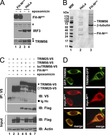
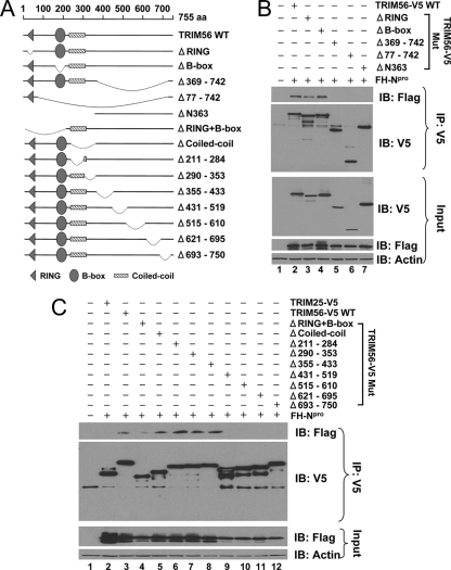
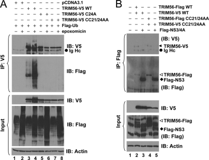
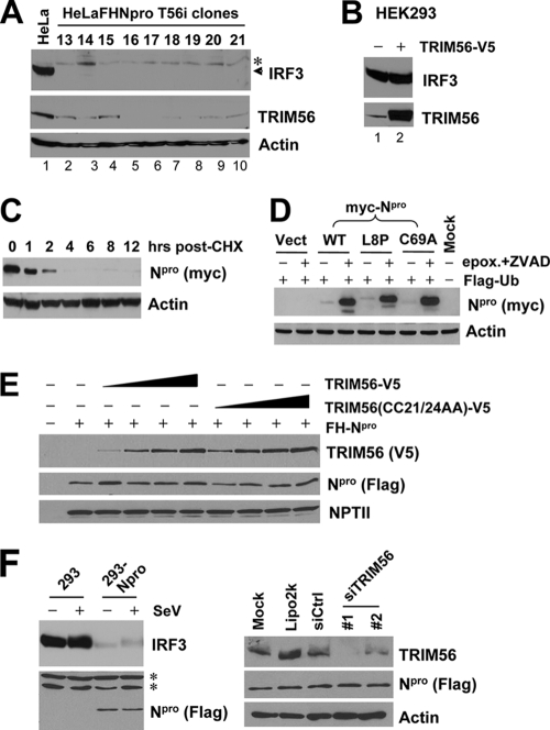
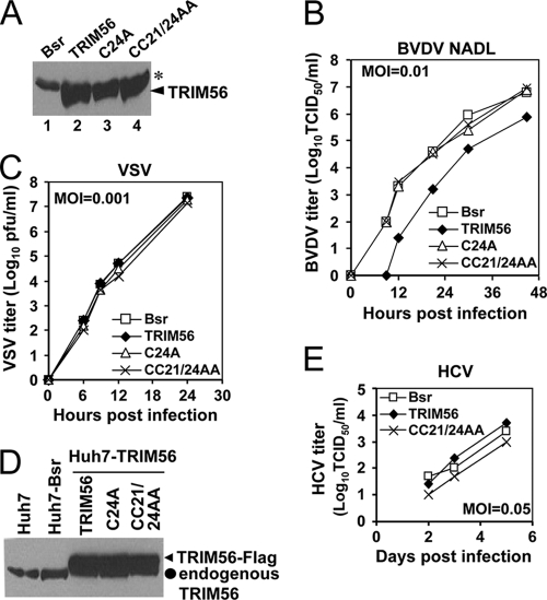
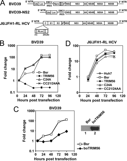
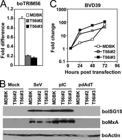
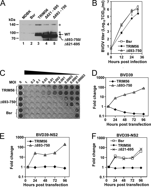
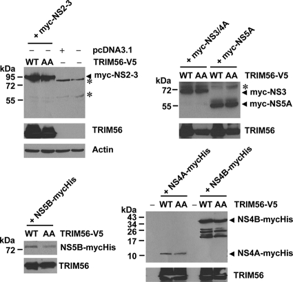
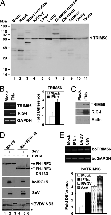
The tripartite motif (TRIM) protein family comprises more than 60 members that have diverse functions in various biological processes. Although a small number of TRIM proteins have been shown to regulate innate immunity, much remains to be learned about the functions of the majority of the TRIM proteins. Here we identify TRIM56 as a cellular protein associated with the N-terminal protease (N(pro)) of bovine viral diarrhea virus (BVDV), a pestiviral interferon antagonist which degrades interferon regulatory factor 3 (IRF3) through the proteasome. We found that TRIM56 was constitutively expressed in most tissues, and its abundance was further upregulated moderately by interferon or virus. The manipulation of TRIM56 abundance did not affect the protein turnover of N(pro) and IRF3. Rather, ectopic expression of TRIM56 substantially impaired, while knockdown of TRIM56 expression greatly enhanced, BVDV replication in cell culture. The antiviral activity of TRIM56 depended on its E3 ubiquitin ligase activity as well as the integrity of its C-terminal region but was not attributed to a general augmentation of the interferon antiviral response. Overexpression of TRIM56 did not inhibit the replication of vesicular stomatitis virus or hepatitis C virus, a virus closely related to BVDV. Together, our data demonstrate that TRIM56 is a novel antiviral host factor that restricts pestivirus infection.


Figures

Fig. 1.
Fig. 1.
Identification of TRIM56 as a protein interaction partner of BVDV Npro. (A) Immunoblot analysis of Flag- and HA-tandem-tagged Npro (FHNpro) (using anti-Flag antibody), IRF3, and TRIM56 expression in HeLa-FHNpro (lanes 1 and 2) and parental HeLa (lanes 3 and 4) cells. Where indicated, cells were treated with epoxomicin (100 nM) overnight prior to cell lysis. Asterisks indicate nonspecific bands, which demonstrate equal sample loading. (B) Protein complexes were purified from control HeLa and HeLa-FHNpro cells by anti-Flag affinity purification and fractionated on SDS-PAGE gels, followed by Coomassie blue staining. The protein bands for TRIM56, β-tubulin, and FH-Npro (identified later by mass spectrometry) are indicated on the right. (C) Co-IP analysis of the interaction of FH-Npro with V5-tagged human TRIM56 (TRIM56) and bovine TRIM56 (boTRIM56) in cotransfected 293FT cells. TRIM25-V5 was used as a negative control for the specificity of the Npro-TRIM56 interaction. Where indicated, cells were treated with epoxomicin overnight prior to cell lysis. Note that epoxomicin had no effect on the Npro-TRIM56/boTRIM56 associations. The input panel shows the immunoblotting of 1/10 of the whole-cell lysates used for IP. Note that ectopically expressed FH-Npro was detected as a doublet in immunoblot analyses of the whole-cell lysates. The upper band may represent monoubiquitinated FH-Npro. IB, immunoblot; Ig Hc, IgG heavy chain. (D) HeLaNpro-25 cells induced for Npro expression were transiently transfected with a vector encoding GFP-TRIM56 (left) or TRIM56-GFP (right) and subsequently fixed for immunostaining of Npro (using anti-Myc antibody). The subcellular localizations of GFP-tagged TRIM56 and Myc-Npro were examined by confocal microscopy.
Fig. 2.
Fig. 2.
The C-terminal portion of TRIM56 is important for its association with Npro. (A) Schematic representation of human TRIM56 protein domains and the individual TRIM56 deletion mutants tested in this study. (B and C) Co-IP analysis of the association of FH-Npro with V5-tagged, WT TRIM56 or the indicated TRIM56 mutant in cotransfected 293FT cells. In C, TRIM25-V5 was used as a negative control for the interaction with FH-Npro.
Fig. 3.
Fig. 3.
TRIM56 is a RING-type E3 Ub ligase and self-associates. (A) HEK293 cells were cotransfected with Flag-Ub, pcDNA3.1, and WT or C24A or CC21/24AA mutant TRIM56-V5 and mock treated or treated with 50 nM epoxomicin. (Top) Cell lysates were subjected to IP with anti-V5 MAb, followed by immunoblot analysis with anti-V5 MAb and anti-Flag M2. (Bottom) Immunoblot analysis of whole-cell lysates (input) for WT and C24A and CC21/24AA mutant TRIM56-V5 (using MAb anti-V5 antibody), Flag-Ub (using rabbit anti-Flag), and actin. (B) Co-IP analysis of the interactions of TRIM56-Flag with TRIM56-V5 (lane 2), TRIM56-V5 with Flag-NS3/4A (lane 3, as a negative control), CC21/24AA TRIM56-Flag with CC21/24AA TRIM56-V5 (lane 4), and CC21/24AA TRIM56-V5 with Flag-NS3/4A (lane 5, as a negative control) in cotransfected 293FT cells. (Top) Cell lysates were immunoprecipitated with anti-Flag, followed by immunoblot analysis with anti-V5 MAb and anti-Flag MAb. (Bottom) Immunoblot analysis of whole-cell lysates (input) for WT and CC21/24AA mutant TRIM56-V5 (using anti-V5 MAb), WT and CC21/24AA mutant TRIM56-Flag, Flag-NS3/4A (using rabbit anti-Flag), and actin.
Fig. 4.
Fig. 4.
TRIM56 does not regulate the protein abundances of IRF3 and BVDV Npro. (A) Knockdown of TRIM56 does not reverse Npro-induced IRF3 degradation. Data shown are for immunoblot analyses of IRF3, TRIM56, and actin in control HeLa cells (lane 1) and HeLa-FHNpro-derived cell clones stably transfected with a TRIM56 shRNA (lanes 2 to 10). The asterisk indicates a nonspecific band. (B) Overexpression of TRIM56 does not reduce IRF3 protein levels in transiently transfected HEK293 cells. (C) Immunoblot analysis of Npro and actin in HeLaNpro-25 cells induced for Npro expression following treatment with cycloheximide (CHX) (75 μg/ml) for the indicated times. (D) Immunoblot analysis of Npro and actin expression in 293FT cells cotransfected with Flag-Ub and Myc-tagged WT or L8P or C69A mutant Npro. Where indicated, cells were treated with epoxomicin (50 nM) plus a pancaspase inhibitor, ZVAD (40 μM). (E) Overexpression of WT or mutant TRIM56 does not regulate the protein level of cotransfected FH-Npro in HEK293 cells. The empty pcDNA3.1 vector was supplemented to keep the total DNA transfected constant. An immunoblot of neomycin phosphotransferase II (NPTII) is shown to demonstrate equal transfection efficiency and sample loading. (F) Knockdown of endogenous TRIM56 does not regulate Npro protein abundance in HEK293 cells stably expressing Flag-Npro (293-Npro). (Left) Immunoblot analysis of IRF3 and Npro in control HEK293 and 293-Npro cells (mock infected or infected with SeV). (Right) Immunoblot analysis of TRIM56, Npro, and actin expression in 293-Npro cells mock transfected or transfected with Lipofectamine alone or Lipofectamine complexed with a negative-control siRNA or with individual siRNAs (siRNAs 1 and 2) specifically targeting TRIM56.
Fig. 5.
Fig. 5.
Ectopically expressed TRIM56 restricts BVDV replication in bovine kidney (MDBK) cells by means of its E3 Ub ligase activity. (A) Immunoblot analysis of ectopically expressed TRIM56 in MDBK cells stably expressing a control vector (Bsr), WT TRIM56, and RING mutant TRIM56 (C24A and CC21/24AA). The asterisk indicates a nonspecific band that demonstrates equal sample loading. (B) Progeny virus production in culture supernatants of MDBK-Bsr, -TRIM56, -C24A, and -CC21/24AA cells at various time points postinfection with BVDV NADL (MOI of 0.01). Data shown are representative of three independently conducted experiments. (C) Progeny virus production in culture supernatants of MDBK-Bsr, -TRIM56, -C24A, and -CC21/24AA cells at various time points postinfection with VSV (MOI of 0.001). Data shown are representative of two independently conducted experiments. (D) Immunoblot analysis of TRIM56 expression (using anti-TRIM56) in parental Huh7 cells and Huh7 cells stably transduced with the control vector (Bsr), WT TRIM56 (TRIM56), or the TRIM56 RING mutants (C24A or CC21/24AA). (E) Progeny virus production in culture supernatants of Huh7-Bsr, -TRIM56, and -CC21/24AA cells at various time points postinfection with HCV JFH1 (MOI of 0.05). At day 1, the infectious progeny virus titers were all below the detection limit (5 TCID50/ml). Data shown are representative of two independently conducted experiments.
Fig. 6.
Fig. 6.
TRIM56 inhibits BVDV infection by targeting intracellular viral RNA replication. (A) Schematic representation of the subgenomic ncpBVDV RNA replicons encoding firefly luciferase, BVD39 (which encodes Npro) (15) and BVD39-NS2 (which does not encode Npro), and the genome-length J6/JFH1-RL HCV replicon encoding Renilla luciferase (32). NTR, nontranslated region. (B) Replication of the BVD39 replicon in transfected MDBK-Bsr, -TRIM56, -C24A, and -CC21/24AA cells. Data shown represent the fold increases in luciferase activity over that present at 6 h (which represents the direct translation of input replicon RNA) in each cell type and are representative of two independently conducted experiments. (C, left) Replication of the BVD39 replicon in MDBK-Bsr cells and MDBK-boTRIM56 cells stably overexpressing boTRIM56. Data shown are representative of two independent experiments. (Right) Immunoblot analysis of ectopically expressed boTRIM56 (using anti-Flag antibody) in MDBK-boTRIM56 cells (lane 2). MDBK-Bsr cells (lane 1) served as a negative control. (D) Replication of the J6/JFH1-RL HCV replicon in transfected Huh7 and Huh7-Bsr, -TRIM56, -C24A, and -CC21/24AA cells. Data shown represent the fold increases in luciferase activity over that present at 6 h in each cell type and are representative of two independent experiments.
Fig. 7.
Fig. 7.
The endogenous bovine TRIM56 restricts BVDV replication in MDBK cells. (A) Real-time RT-PCR analysis of boTRIM56 mRNA expression in parental MDBK cells and two MDBK cell clones stably transfected with a boTRIM56 shRNA (T56i#2 and T56i#3). (B) Effect of the stable knockdown of boTRIM56 in MDBK cells on viral induction of the type I IFN response. Parental MDBK and MDBK-T56i #2 and -T56i#3 cells were mock treated, infected with SeV, or transfected with poly(I:C) (pIC) or poly(dA:dT) (pdAdT). Cells were lysed for immunoblot analysis of bovine ISG15, MxA, and actin expression. Note that MDBK cells did not respond to transfected poly(dA:dT) to induce ISG15 and MxA expression. (C) Replication of the BVD39 replicon in MDBK and MDBK-T56i #2 and -T56i#3 cells. Data shown are representative of two independent experiments.
Fig. 8.
Fig. 8.
The C-terminal structural integrity is important for TRIM56's antiviral function against BVDV. (A) Immunoblot detection of ectopically expressed TRIM56 in parental MDBK cells (lane 1) and MDBK cells stably expressing the Bsr vector (lane 2), WT TRIM56 (lane 3), or the Δ621-695 (lane 4) and Δ693-750 (lane 5) mutant TRIM56 proteins. Asterisks indicate nonspecific bands. (B) Progeny BVDV titers in culture supernatants of MDBK-Bsr, MDBK-TRIM56, and MDBK-Δ693-750 mutant cells at various time points postinfection with cp BVDV NADL (MOI of 0.005). Data shown are representative of three independent experiments. (C) Duplicate wells of MDBK-TRIM56, MDBK-Δ693-750 mutant, and MDBK-Bsr cells grown in a 96-well plate were infected with increasing MOIs (from right to left, MOIs of 0.00003 through 1) of the cp BVDV NADL for 70 h prior to cell fixation and crystal violet staining. The very left column shows uninfected cells. The image shown is representative of two independent experiments. (D and E) Replication of the BVD39 (D) and BVD39-NS2 (E) replicons in MDBK-TRIM56 and MDBK-Δ693-750 mutant cells. Data shown are representative of three independent experiments. (F) Replication of the BVD39-NS2 replicon in MDBK-Bsr, MDBK-TRIM56, and MDBK-Δ621-695 mutant cells. Data shown are representative of three independent experiments.
Fig. 9.
Fig. 9.
TRIM56 does not promote the degradation of BVDV NS proteins. 293FT cells grown in 6-well plates were mock transfected or cotransfected with 0.25 μg of the indicated Myc- or Myc-6×His-tagged ncpBVDV NS protein construct and 1.5 μg of the V5-tagged TRIM56 (WT or the CC21/24AA mutant [AA]) vector. Cells were lysed and subjected to immunoblot analysis of the expression of BVDV NS proteins (using anti-Myc antibody) and TRIM56 (using anti-V5 antibody). Asterisks indicate nonspecific bands.
Fig. 10.
Fig. 10.
TRIM56 is ubiquitously expressed, and its expression is further upregulated by virus or IFN-α. (A) Expression of the TRIM56 protein in various human tissues detected by anti-TRIM56 antibody using Human Tissues INSTA-Blot (Calbiochem). (B) Semiquantitative RT-PCR (left) and real-time PCR (right) assessments of TRIM56 mRNA abundance in HeLa cells mock treated or stimulated with 500 U/ml of IFN-α for 16 h. RIG-I was used as a positive control for IFN treatment (left). (C) Immunoblot analysis of TRIM56 and RIG-I expression in THP1 cells mock treated or stimulated with IFN-α. Similar results were obtained with HeLa cells (data not shown). (D) Stable retroviral transduction of IRF3, but not that of IRF3 with an N-terminal 133-aa deletion (DN133), renders MDBK cells able to upregulate boISG15 expression following BVDV infection. Cells were mock treated (lanes 1 and 4) or infected with SeV (lanes 2 and 5) or with BVDV (lanes 3 and 6) for 12 h prior to cell lysis and immunoblot analysis of the ectopically expressed, Flag- and HA-tandem-tagged human IRF3 (FH-IRF3) or IRF3 DN133 (FH-IRF3DN133) (using anti-Flag antibody), bovine ISG15, SeV, and BVDV NS3. The asterisk indicates a nonspecific band, which demonstrates equal protein sample loading. (E) Semiquantitative RT-PCR (top) and real-time RT-PCR (bottom) analyses of boTRIM56 mRNA abundance in BK-F3 cells mock treated, stimulated with 500 U/ml of IFN-α, or infected with cp BVDV NADL (MOI of 0.1) or SeV (50 HAU/ml) for 16 h.

References

    1. Barr S. D., Smiley J. R., Bushman F. D. 2008. The interferon response inhibits HIV particle production by induction of TRIM22. PLoS Pathog. 4:e1000007. - PMC - PubMed
    1. Beard M. R., et al. 1999. An infectious molecular clone of a Japanese genotype 1b hepatitis C virus. Hepatology 30:316–324 - PubMed
    1. Carthagena L., et al. 2009. Human TRIM gene expression in response to interferons. PLoS One 4:e4894. - PMC - PubMed
    1. Chan M. W., et al. 2005. Hypermethylation of 18S and 28S ribosomal DNAs predicts progression-free survival in patients with ovarian cancer. Clin. Cancer Res. 11:7376–7383 - PubMed
    1. Chen Z., et al. 2007. Ubiquitination and proteasomal degradation of interferon regulatory factor-3 induced by Npro from a cytopathic bovine viral diarrhea virus. Virology 366:277–292 - PMC - PubMed

Publication types

LinkOut - more resources