. 2011 Feb 2;31(5):1746-51.
doi: 10.1523/JNEUROSCI.5704-10.2011.

Selective toxicity by HDAC3 in neurons: regulation by Akt and GSK3beta

Affiliations

Selective toxicity by HDAC3 in neurons: regulation by Akt and GSK3beta

Farah H Bardai et al. J Neurosci. .

Abstract

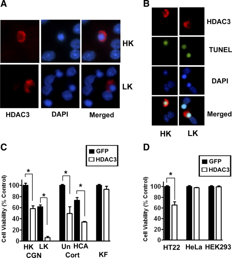
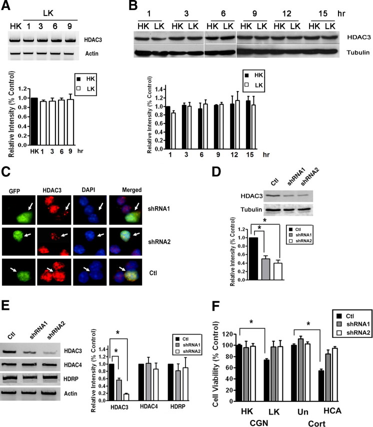
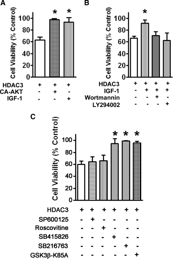
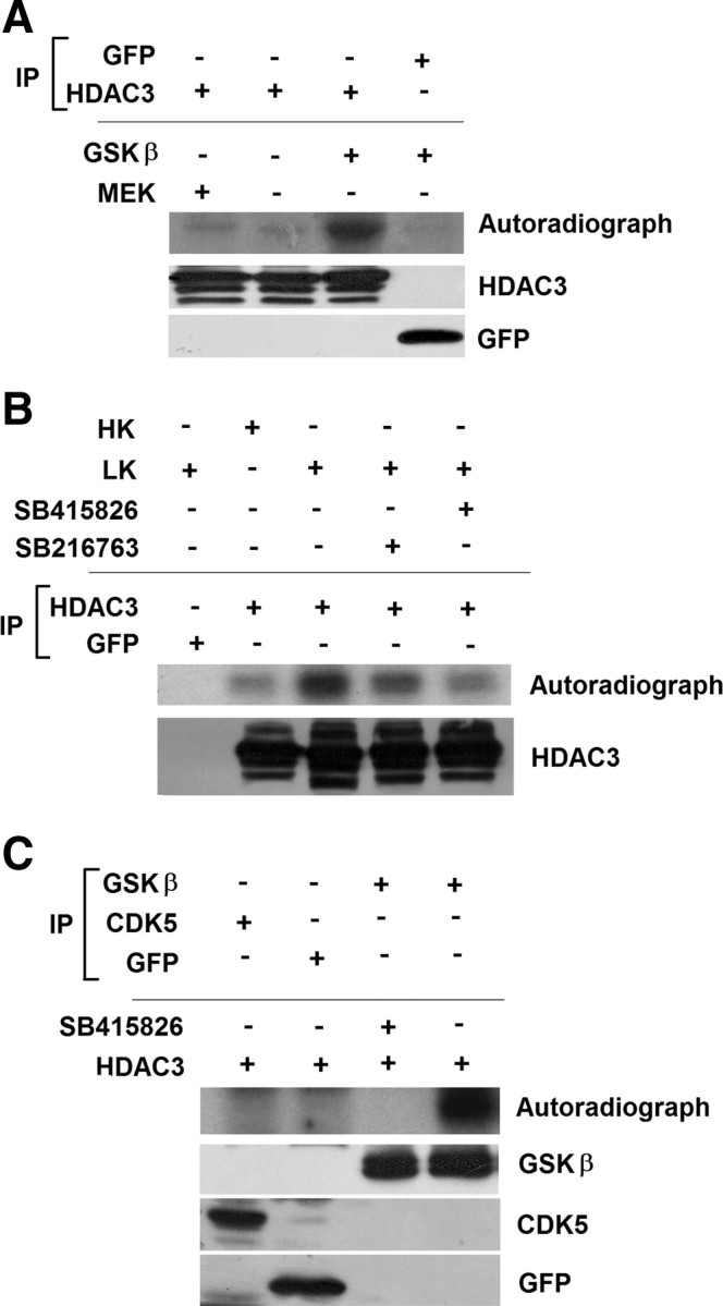
Although it is well established that pharmacological inhibitors of classical histone deacetylases (HDACs) are protective in various in vivo models of neurodegenerative disease, the identity of the neurotoxic HDAC(s) that these inhibitors target to exert their protective effects has not been resolved. We find that HDAC3 is a protein with strong neurotoxic activity. Forced expression of HDAC3 induces death of otherwise healthy rat cerebellar granule neurons, whereas shRNA-mediated suppression of its expression protects against low-potassium-induced neuronal death. Forced expression of HDAC3 also promotes the death of rat cortical neurons and hippocampally derived HT22 cells, but has no effect on the viability of primary kidney fibroblasts or the HEK293 and HeLa cell lines. This suggests that the toxic effect of HDAC3 is cell selective and that neurons are sensitive to it. Neurotoxicity by HDAC3 is inhibited by treatment with IGF-1 as well as by the expression of a constitutively active form of Akt, an essential mediator of IGF-1 signaling. Protection against HDAC3-induced neurotoxicity is also achieved by the inhibition of GSK3β, a kinase inhibited by Akt that is widely implicated in the promotion of neurodegeneration in experimental models and in human pathologies. HDAC3 is directly phosphorylated by GSK3β, suggesting that the neuronal death-promoting action of GSK3β could be mediated through HDAC3 phosphorylation. In addition to demonstrating that HDAC3 has neurotoxic effects, our study identifies it as a downstream target of GSK3β.


Figures

Figure 1.
Figure 1.
Forced expression of HDAC3 kills neurons selectively. Primary neuronal and non-neuronal cells and cell lines were transfected with plasmids encoding HDAC3-Flag or GFP. Effect of elevated HDAC3 expression on cell viability was evaluated. A, Immunofluorescence images of CGNs transfected with HDAC3-Flag and later switched to HK or LK medium for 24 h. B, TUNEL staining of HDAC3-Flag transfected CGNs after treatment with HK or LK for 24 h. C, Quantification of viability in primary cultures. CGNs, cortical neurons (Cort), and kidney fibroblasts (KF) were transfected with HDAC3 or GFP-expressing plasmids. Eight hours after transfection, CGNs were switched to either HK or LK medium and cortical neurons were either left untreated (Un) or treated with HCA. Kidney fibroblasts were left without additional treatments. D, Quantification of viability in cell lines. HT22, HeLa, and HEK293 cells were transfected with HDAC3 or GFP. Viability was quantified 32 h after transfection and compared with control cultures transfected with GFP. *p < 0.001.
Figure 2.
Figure 2.
Suppression of HDAC3 expression protects neurons. A, CGNs were treated with LK for 1, 3, 6, or 9 h. Control cultures received HK for 9 h (HK). Total RNA was extracted and RT-PCR performed using specific primers for HDAC3. Actin served as loading control. The lower panel shows the densitometric analysis of the RT-PCR data from three separate experiments. B, CGNs were treated with HK or LK medium for 1, 3, 6, 9, 12, or 15 h. Expression of HDAC3 protein was analyzed by Western blotting. The blot was reprobed with a tubulin antibody. Densitometric analysis of the bands from different experiments was performed and normalized to tubulin. C, CGNs were cotransfected with plasmids expressing GFP with control shRNA (Ctl), shRNA-1, or shRNA-2 for 72 h. The cells were costained with GFP and HDAC3 antibodies and nuclear morphology visualized by DAPI staining. Arrows point to successfully transfected cells (GFP-positive). Neurons receiving either shRNA construct show reduced HDAC3 immunostaining whereas cells cotransfected with the control shRNA show similar HDAC3 staining as untransfected cells. D, E, Lysates from HT22 cells transfected with control shRNA, shRNA-1, or shRNA-2 were analyzed by Western blotting or RT-PCR. D, Western blot results using HDAC3 and tubulin antibodies. Densitometric analysis of the bands was performed and normalized to tubulin. E, Results of RT-PCR analysis of HDAC3, HDAC4, HDRP, and actin (loading control) expression. Also included is densitometric analysis of the data from three different experiments normalized to actin. F, CGNs and cortical neurons (Cort) were transfected with plasmids expressing control shRNA, shRNA-1, or shRNA-2 and cell viability quantified as described in Materials and Methods. After transfection, CGNs were treated with HK or LK for 24 h whereas cortical neurons were either untreated (Un) or treated with HCA for 20 h. Cell viability is normalized to survival in cultures transfected with control shRNA and treated in HK (CGN) or untreated (Cort). *p < 0.05.
Figure 3.
Figure 3.
HDAC3-induced neurotoxicity is inhibited by IGF-1 and activation of the PI-3 kinase–Akt signaling pathway. A, CGNs were transfected with HDAC3-Flag alone or in combination with a plasmid expressing HA-tagged constitutively active Akt (CA-Akt-HA). The transfected cultures were then treated with no additives or with IGF-1. B, CGNs were transfected with HDAC3-Flag and then treated with no additives or with IGF-1, IGF-1, and wortmannin, or IGF-1 and LY294002. Eight hours later, the cultures were switched to serum-free medium containing inhibitors, and cell viability assessed 24 h later. C, CGNs were transfected with HDAC3-Flag and treated with HK medium containing either no additives, supplemented with JNK inhibitor (SP600125), CDK inhibitor (roscovitine), GSK3 inhibitor (SB415826 or SB216763), or cotransfected with a dominant-negative form of GSK3β (GSK3β-K85A). Cell viability was assessed 24 h after switching to HK medium. Viability in all cases was normalized to control cultures, which were transfected with GFP and treated with HK medium. *p < 0.01.
Figure 4.
Figure 4.
HDAC3 is directly phosphorylated by GSK3β. A, Lysates from HEK293 cells transfected with either GFP or HDAC3 were immunoprecipitated (IP) using either GFP or Flag antibody. The immunoprecipitated proteins were used in an in vitro kinase assay with or without active GSK3β or with active MEK. The reaction mixture was subjected to PAGE and phosphorylation evaluated by autoradiography. Immunoblots show that HDAC3-Flag and GFP were immunoprecipitated in the samples used for the kinase assay. B, CGN cultures were metabolically labeled with [32P] orthophospate in HK, LK, or LK media with GSK3 inhibitors for 3 h. Lysates from the cultures were immunoprecipitated using GFP or HDAC3 antibody and the extent of phosphorylation evaluated by PAGE and autoradiography. The bottom shows that HDAC3 was immunoprecipitated in the samples used for the kinase assay. C, Equal amounts of GSK3β, CDK5, or GFP immunoprecipitated from CGNs treated with LK for 6 h were combined with HDAC3 in an in vitro kinase assays with or without a GSK3 inhibitor as indicated. HDAC3 phosphorylation was evaluated by PAGE of the reaction mixture and autoradiography. Bottom, Similar amounts of GFP, GSK3β, and CDK5 were used in the assay.

References

    1. Bates EA, Victor M, Jones AK, Shi Y, Hart AC. Differential contributions of Caenorhabditis elegans histone deacetylases to huntingtin polyglutamine toxicity. J Neurosci. 2006;26:2830–2838. - PMC - PubMed
    1. Bhat RV, Budd Haeberlein SL, Avila J. Glycogen synthase kinase 3: a drug target for CNS therapies. J Neurochem. 2004;89:1313–1317. - PubMed
    1. Borsello T, Forloni G. JNK signalling: a possible target to prevent neurodegeneration. Curr Pharm Des. 2007;13:1875–1886. - PubMed
    1. Boyault C, Zhang Y, Fritah S, Caron C, Gilquin B, Kwon SH, Garrido C, Yao TP, Vourc'h C, Matthias P, Khochbin S. HDAC6 controls major cell response pathways to cytotoxic accumulation of protein aggregates. Genes Dev. 2007;21:2172–2181. - PMC - PubMed
    1. Camins A, Verdaguer E, Folch J, Canudas AM, Pallàs M. The role of CDK5/P25 formation/inhibition in neurodegeneration. Drug News Perspect. 2006;19:453–460. - PubMed

Publication types

MeSH terms

LinkOut - more resources