. 2011 Jan 26:5:3.
doi: 10.3389/fnana.2011.00003. eCollection 2011.

Cladistic analysis of olfactory and vomeronasal systems

Affiliations

Cladistic analysis of olfactory and vomeronasal systems

Isabel Ubeda-Bañon et al. Front Neuroanat. .

Abstract

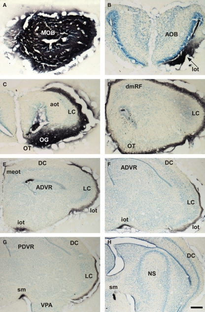
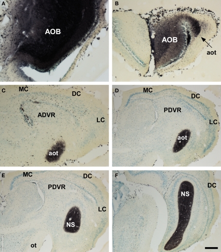
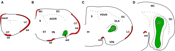
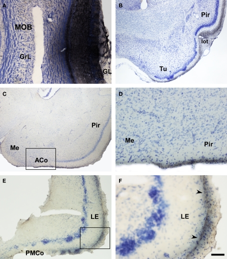
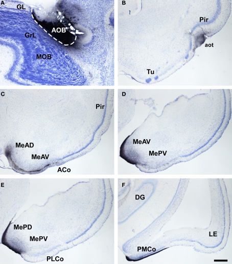
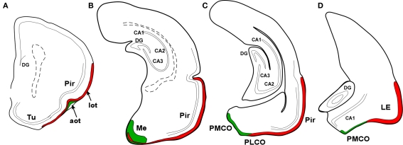
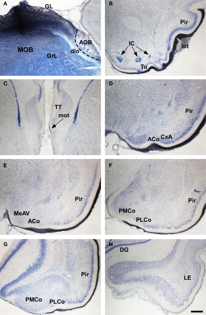
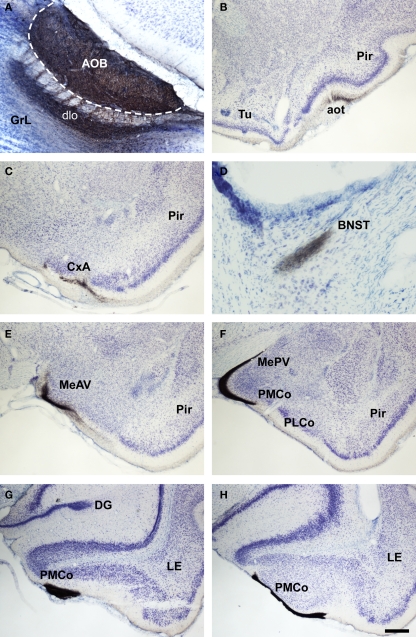
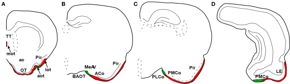
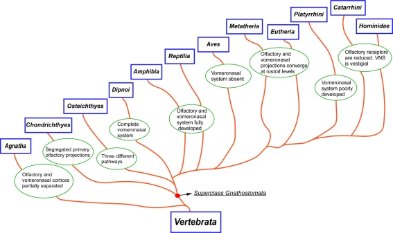
Most tetrapods possess two nasal organs for detecting chemicals in their environment, which are the sensory detectors of the olfactory and vomeronasal systems. The seventies' view that the olfactory system was only devoted to sense volatiles, whereas the vomeronasal system was exclusively specialized for pheromone detection was challenged by accumulating data showing deep anatomical and functional interrelationships between both systems. In addition, the assumption that the vomeronasal system appeared as an adaptation to terrestrial life is being questioned as well. The aim of the present work is to use a comparative strategy to gain insight in our understanding of the evolution of chemical "cortex." We have analyzed the organization of the olfactory and vomeronasal cortices of reptiles, marsupials, and placental mammals and we have compared our findings with data from other taxa in order to better understand the evolutionary history of the nasal sensory systems in vertebrates. The olfactory and vomeronsasal cortices have been re-investigated in garter snakes (Thamnophis sirtalis), short-tailed opossums (Monodelphis domestica), and rats (Rattus norvegicus) by tracing the efferents of the main and accessory olfactory bulbs using injections of neuroanatomical anterograde tracers (dextran-amines). In snakes, the medial olfactory tract is quite evident, whereas the main vomeronasal-recipient structure, the nucleus sphaericus is a folded cortical-like structure, located at the caudal edge of the amygdala. In marsupials, which are acallosal mammals, the rhinal fissure is relatively dorsal and the olfactory and vomeronasal cortices relatively expanded. Placental mammals, like marsupials, show partially overlapping olfactory and vomeronasal projections in the rostral basal telencephalon. These data raise the interesting question of how the telencephalon has been re-organized in different groups according to the biological relevance of chemical senses.

Keywords: amygdala; cortex; evolution; olfaction; olfactory bulb; vomeronasal.


Figures

Figure 1
Figure 1
Bright field microscopic images from rostral to caudal of coronal sections of one brain hemisphere showing biotinylated dextran-amine labeling Nissl-counterstained in the snake (Thamnophis sirtalis) olfactory cortices after one injection in the main olfactory bulb (MOB). (A) injection site in the MOB. (B–H) labeling in olfactory-recipient areas of the telencephalon. Scale bar A–D = 267; E–H = 400 μm.
Figure 2
Figure 2
Bright field microscopic images from rostral to caudal of coronal sections of one brain hemisphere showing biotinylated dextran-amine labeling Nissl-counterstained in the snake (Thamnophis sirtalis) vomeronasal amygdala after an injection in the accessory olfactory bulb (AOB). (A) injection site. (B–F) labeling in vomeronasal-recipient areas. Scale bar A = 160; B–C = 267; D–E = 400; F = 533 μm.
Figure 3
Figure 3
Schematic representation of main (red) and accessory (green) projections from rostral to caudal (A-D) areas in the snake (Thamnophis sirtalis) olfactory – and vomeronasal-recipient areas.
Figure 4
Figure 4
Bright field microscopic images from rostral to caudal of coronal sections of one brain hemisphere showing biotinylated dextran-amine labeling Nissl-counterstained in the opossum (Monodelphis domestica) olfactory cortices after an injection in the main olfactory bulb (MOB). (A) injection site. (B–F) labeling in olfactory-recipient areas. Scale bar A, D, and F = 160; B, C, and E = 800 μm.
Figure 5
Figure 5
Bright field microscope images from rostral to caudal of coronal (A is sagittal) sections of one brain hemisphere showing biotinylated dextran-amine labeling Nissl-counterstained in the opossum (Monodelphis domestica) vomeronasal cortices after an injection in the accessory olfactory bulb (AOB). (A) injection site. (B) labeling in vomeronasal-recipient areas. Scale bar A = 400; B–F = 800 μm.
Figure 6
Figure 6
Schematic representation of main (red) and accessory (green) projections from rostral to caudal (A-D) areas in the opossum (Monodelphis domestica) olfactory and vomeronasal cortices.
Figure 7
Figure 7
Bright field microscopic images from rostral to caudal of coronal (A is sagittal) sections of one brain hemisphere showing biotinylated dextran-amine labeling Nissl-counterstained in the rat (Rattus norvegicus) olfactory cortices after an injection in the main olfactory bulb (MOB). (A) injection site. (B–H) labeling in olfactory-recipient areas. Scale bar A–B and DH = 800; C = 400 μm.
Figure 8
Figure 8
Bright field microscopic images from rostral to caudal of coronal (A is sagittal) sections of one brain hemisphere showing biotinylated dextran-amine labeling Nissl-counterstained in the rat (Rattus norvegicus) vomeronasal cortices after an injection in the accessory olfactory bulb (AOB). (A) injection site. (B–H) labeling in vomeronasal-recipient areas. Scale bar A = 400; B–C and E–H = 800; D = 160 μm.
Figure 9
Figure 9
Schematic representation of main (red) and accessory (green) projections from rostral to caudal (A-D) areas in the rat (Rattus norvegicus) olfactory and vomeronasal cortices.
Figure 10
Figure 10
Cladogram illustrating the main taxa analyzed in the present report and the main changes in the olfactory and vomeronasal systems occurred during evolution.

Similar articles

Cited by

References

    1. Abellan A., Legaz I., Vernier B., Retaux S., Medina L. (2009). Olfactory and amygdalar structures of the chicken ventral pallium based on the combinatorial expression patterns of LIM and other developmental regulatory genes. Comp. J. Neurol. 516, 166–186 - PubMed
    1. Abolmaali N. D., Kuhnau D., Knecht M., Kohler K., Huttenbrink K. B., Hummel T. (2001). Imaging of the human vomeronasal duct. Chem. Senses 26, 35–3910.1093/chemse/26.1.35 - DOI - PubMed
    1. Ache B. W., Young J. M. (2005). Olfaction: diverse species, conserved principles. Neuron 48, 417–43010.1016/j.neuron.2005.10.022 - DOI - PubMed
    1. Adey W. R. (1953). An experimental study of the central olfactory connexions in a marsupial (Trichosurus Vulpecula). Brain 76, 311–33010.1093/brain/76.2.311 - DOI - PubMed
    1. Alonso J. R., Covenas R., Lara J., Arevalo R., de Leon M., Aijon J. (1989). Tyrosine hydroxylase immunoreactivity in a subpopulation of granule cells in the olfactory bulb of teleost fish. Brain Behav. Evol. 34, 318–324 - PubMed

LinkOut - more resources