. 2011 May 20;6(5):492-501.
doi: 10.1021/cb100410m. Epub 2011 Feb 22.

Discovery of a potential allosteric ligand binding site in CDK2

Affiliations

Discovery of a potential allosteric ligand binding site in CDK2

Stephane Betzi et al. ACS Chem Biol. .

Abstract

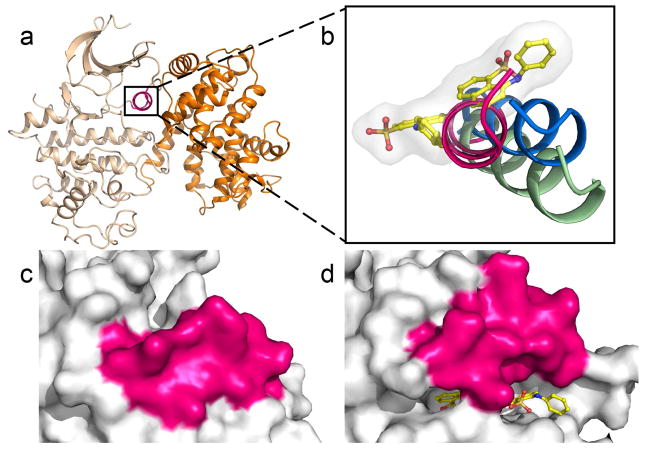
Cyclin-dependent kinases (CDKs) are key regulatory enzymes in cell cycle progression and transcription. Aberrant activity of CDKs has been implicated in a number of medical conditions, and numerous small molecule CDK inhibitors have been reported as potential drug leads. However, these inhibitors exclusively bind to the ATP site, which is largely conserved among protein kinases, and clinical trials have not resulted in viable drug candidates, attributed in part to the lack of target selectivity. CDKs are unique among protein kinases, as their functionality strictly depends on association with their partner proteins, the cyclins. In an effort to identify potential target sites for disruption of the CDK-cyclin interaction, we probed the extrinsic fluorophore 8-anilino-1-naphthalene sulfonate (ANS) with human CDK2 and cyclin A using fluorescence spectroscopy and protein crystallography. ANS interacts with free CDK2 in a saturation-dependent manner with an apparent K(d) of 37 μM, and cyclin A displaced ANS from CDK2 with an EC(50) value of 0.6 μM. Co-crystal structures with ANS alone and in ternary complex with ATP site-directed inhibitors revealed two ANS molecules bound adjacent to one another, away from the ATP site, in a large pocket that extends from the DFG region above the C-helix. Binding of ANS is accompanied by substantial structural changes in CDK2, resulting in a C-helix conformation that is incompatible for cyclin A association. These findings indicate the potential of the ANS binding pocket as a new target site for allosteric inhibitors disrupting the interaction of CDKs and cyclins.


Figures

Figure 1
Figure 1
ANS and the ATP site-directed inhibitors JWS648 (IC50 = 5.9 μM) and SU9516 (IC50 = 0.13 μM).
Figure 2
Figure 2
Fluorescence measurements of CDK2-ANS interaction, the effect of selected CDK2 ligands, and the efficency of ANS interaction with other protein kinases. a) Emission spectra of CDK2 (1.6 μM) were recorded for increasing concentrations of ANS. Fitting the emission maximum at 460 nm to equation (3) yielded an apparent Kd of 37 μM (inset). b) Emission spectra for the CDK2-ANS complex were recorded at 50 μM ANS and 1.6 μM CDK2 in the presence of increasing concentrations of cyclin A2. Cyclin A2 quenched the fluorescence yield in a dose-dependent manner, indicating the displacement of ANS with an EC50 value of 0.6 μM (inset). c) Spectra were recorded for 50 μM ANS and 1.6 μM CDK2 in the presence of small molecule ligands and the fluorescence maximum at 460 nm analyzed as a function of compound concentration. ATP site-directed ligands exhibited differential ANS displacement potential: SU9515 (■) readily quenched the fluorescence signal (EC50 = 0.3 μM), while concentrations as high as 300 μM of JWS648 (▼) and ATP (●) only marginally affected the fluorescence yield. Note that the displacement potential of SU9515 decreased in the presence of 50 μM JWS648 (□) to EC50 = 1.2 μM. Spectra for a-c were recorded with emission and excitation slit widths 5/5. d) ANS titrations were also performed for Aurora A (5.8 μM) (■), Rock1 (4.3 μM) (◇) and Akt 1 (4.1 μM) (▼) in parallel with CDK2 (1.5 μM) (●). Plotted is the maximum fluorescence yield observed per μM respective kinase in the presence of increasing ANS concentrations. Spectra for d were recorded with slit widths 10/10. Data were fitted to equation (3) yielding Kd values of 37 μM for CDK2, 54 μM for Aurora A, 450 μM for Akt and 1900 μM for Rock 1.
Figure 3
Figure 3
Molecular mode of action of ANS binding to CDK2. a) Two ANS molecules (yellow) bind adjacent to one another, away from the ATP site (hinge region shown in cyan) and in the vicinity of the C-helix (magenta). Displayed in blue is the 2Fo-Fc electron density, contoured at 1σ around both ANS molecules. The Fo-Fc electron density map with the ANS molecules omitted during refinement is shown in Fig. S7a. b) Schematic representation of potential hydrogen bonding and van der Waals (hydrophobic) interactions between ANS and enzyme residues. c) Stereoview of binding interactions within the ANS pocket. Black and green dotted lines indicate hydrogen bonding and hydrophobic interactions, respectively. Water molecules are shown as blue spheres.
Figure 4
Figure 4
Binding of ANS induces large conformational changes in the C-helix. a) Overall view of the CDK2-cyclin A complex structure (PDB entry 2CCI) (45). CDK2 is shown in wheat, the C-helix in magenta, and cyclin A in orange. b) Superimposition reveals the distinct conformation of the C-helix in the CDK2-ANS complex (green) and in free CDK2 (blue) as compared to the CDK2-cyclin A complex (magenta). Surface representations of the C-helix and surrounding residues for free CDK2 (c) and the CDK2-ANS complex (d) reveal the partial opening of the ANS pocket towards solvent.
Figure 5
Figure 5
Allosteric nature of the ANS binding pocket. Crystal structures of ternary complexes with ATP site-directed inhibitors were determined for CDK2-ANS-JWS648 (a) and CDK2-ANS-SU9516 (b). The 2Fo-Fc electron density maps contoured at 1σ around the respective ligands are displayed in blue, with inhibitors shown in orange and ANS in yellow. The Fo-Fc electron density maps with the ligands omitted during refinement are presented in the supplemental material (Fig. S7). (c) and (d) represent stereo views of the binding interactions of ligands in the CDK2-ANS-JWS648 and CDK2-ANS-SU9516 complexes, respectively.
Figure 6
Figure 6
Comparison of the ANS pocket in CDK2 with allosteric sites in other protein kinases. a) In CDK2, the allosteric site extends away from the DFG motif (green) and above the C-helix (magenta). In contrast, the allosteric sites in the Abl-Imatinib complex (b, PDB entry 2HYY) (19), the MEK1-PD318088-ATP complex (c, PDB entry 1S9J) (20) and the P38-BIRB796 complex (d, PDB entry 1KV2) (21) extend along the DFG region so that ligands are underneath the C-helix. Allosteric ligands are shown in yellow, ATP site-binders in orange, and the hinge region in cyan.
Figure 7
Figure 7
Residues comprising the ANS binding pocket are largely conserved among CDKs. Pairwise sequence alignments were performed for CDK2 and CDKs1-9, and their overall sequence similarity and identity values are indicated. Identical residues are highlighted in grey, with conservative and non-conservative substitutions in blue and red, respectively.
Figure 8
Figure 8
Potential of the ANS pocket for the discovery of novel CDK2 inhibitors. a) Based on the binding interactions of ANS with CDK2, potential allosteric inhibitors spanning both ANS sites could be designed by attaching aryl groups via rotatable linkers to the 7-position of the naphthalene. Introduction of polar moieties at the 4-position could serve to increase solubility, and the sulfonate group could be replaced by a sulfonamide to improve pharmacological properties. b) HTS using the CDK2-ANS fluorescence assay, in which compounds that compete for binding to the ANS site induce quenching of the fluorescence signal, could lead to the discovery of new chemical scaffolds with allosteric binding potential. The assay could be performed in the presence of ATP site-directed inhibitors such as JWS648 to protect the CDK2-ANS complex from compounds which indirectly affect the ANS pocket.
Scheme 1
Scheme 1
Synthesis of JWS648

Similar articles

Cited by

References

    1. Hall M, Peters G. Genetic alterations of cyclins, cyclin-dependent kinases, and Cdk inhibitors in human cancer. Adv Cancer Res. 1996;68:67–108. - PubMed
    1. Johnson LN. Protein kinase inhibitors: contributions from structure to clinical compounds. Q Rev Biophys. 2009;42:1–40. - PubMed
    1. Zhang J, Yang PL, Gray NS. Targeting cancer with small molecule kinase inhibitors. Nat Rev Cancer. 2009;9:28–39. - PubMed
    1. Bain J, Plater L, Elliott M, Shpiro N, Hastie CJ, McLauchlan H, Klevernic I, Arthur JS, Alessi DR, Cohen P. The selectivity of protein kinase inhibitors: a further update. Biochem J. 2007;408:297–315. - PMC - PubMed
    1. Sherr CJ. Cancer cell cycles. Science. 1996;274:1672–1677. - PubMed

Publication types