. 2011 Aug;20(8):1451-64.
doi: 10.1089/scd.2010.0461. Epub 2011 Mar 23.

Detection, characterization, and spontaneous differentiation in vitro of very small embryonic-like putative stem cells in adult mammalian ovary

Affiliations

Detection, characterization, and spontaneous differentiation in vitro of very small embryonic-like putative stem cells in adult mammalian ovary

Seema Parte et al. Stem Cells Dev. 2011 Aug.

Retraction in

Abstract

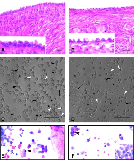
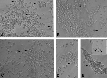
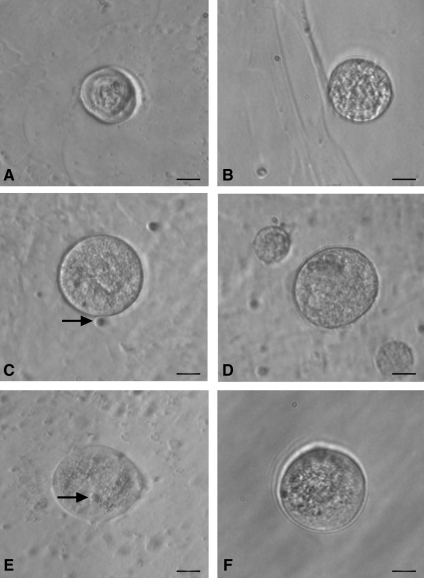
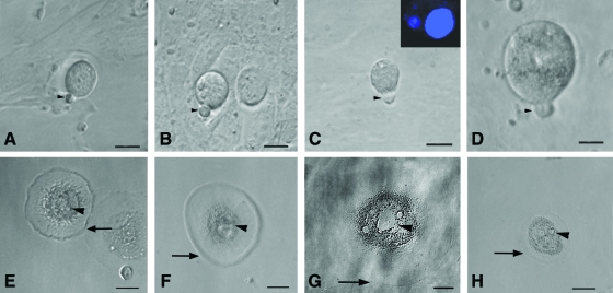
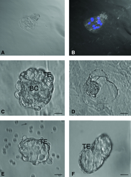
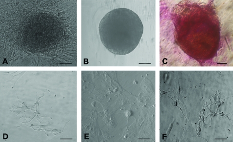
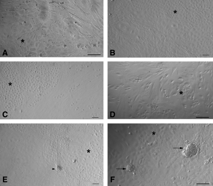
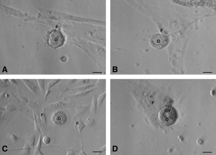
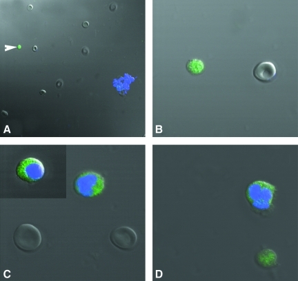
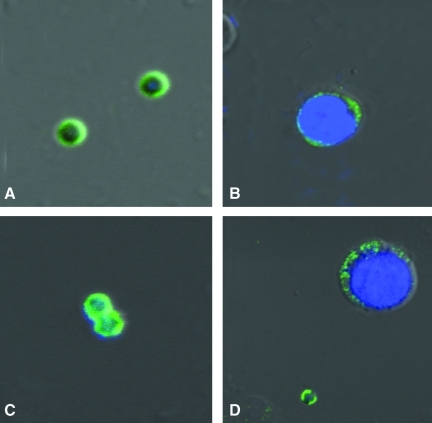
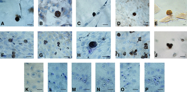
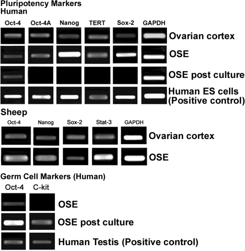
The present study was undertaken to detect, characterize, and study differentiation potential of stem cells in adult rabbit, sheep, monkey, and menopausal human ovarian surface epithelium (OSE). Two distinct populations of putative stem cells (PSCs) of variable size were detected in scraped OSE, one being smaller and other similar in size to the surrounding red blood cells in the scraped OSE. The smaller 1-3 μm very small embryonic-like PSCs were pluripotent in nature with nuclear Oct-4 and cell surface SSEA-4, whereas the bigger 4-7 μm cells with cytoplasmic localization of Oct-4 and minimal expression of SSEA-4 were possibly the tissue committed progenitor stem cells. Pluripotent gene transcripts of Oct-4, Oct-4A, Nanog, Sox-2, TERT, and Stat-3 in human and sheep OSE were detected by reverse transcriptase-polymerase chain reaction. The PSCs underwent spontaneous differentiation into oocyte-like structures, parthenote-like structures, embryoid body-like structures, cells with neuronal-like phenotype, and embryonic stem cell-like colonies, whereas the epithelial cells transformed into mesenchymal phenotype by epithelial-mesenchymal transition in 3 weeks of OSE culture. Germ cell markers like c-Kit, DAZL, GDF-9, VASA, and ZP4 were immuno-localized in oocyte-like structures. In conclusion, as opposed to the existing view of OSE being a bipotent source of oocytes and granulosa cells, mammalian ovaries harbor distinct very small embryonic-like PSCs and tissue committed progenitor stem cells population that have the potential to develop into oocyte-like structures in vitro, whereas mesenchymal fibroblasts appear to form supporting granulosa-like somatic cells. Research at the single-cell level, including complete gene expression profiling, is required to further confirm whether postnatal oogenesis is a conserved phenomenon in adult mammals.


Figures

FIG. 1.
FIG. 1.
H&E-stained sections of menopausal human and adult sheep ovarian cortex (A, B). Note the presence of a continuous OSE in human ovary comprising of cuboidal epithelial cells. OSE was discontinuous with a single layer of cuboidal cells in sheep ovary. Lower panel depicts the presence of spherical stem cells in scraped OSE from human (C) and sheep (D) ovaries, under Hoffman optics. Besides RBCs (asterix) and epithelial cells (arrow), spherical stem cells of 2 distinct sizes viz. small (white arrowhead) and those similar in size to RBCs (black arrowhead) are observed. Note the difference in size of human and sheep RBCs. H&E-stained OSE smears of human (E) and sheep (F) show the presence of epithelial cells with abundant cytoplasm, RBCs, and putative stem cells (PSC) with dark stained nuclei (arrowhead) and minimal cytoplasm. Scale bars=20 μm. OSE, ovarian surface epithelium; H&E, hematoxylin and eosin; RBC, red blood cell.
FIG. 2.
FIG. 2.
PSCs interspersed with RBC and clusters of epithelial cells just after scraping ovarian surface in different mammalian species: (A) rabbit, (B) sheep, (C) monkey, and (D, E) human. Note the presence of small, round, and dark PSCs (arrow head) that are easily distinguished from the relatively bigger, biconcave, and disc-like RBC. The epithelial cells are present in clusters and at places the PSCs are trapped in these clusters. Inset in (E) shows obvious dimensions and size difference between RBC and PSC. Scale bar=50 μm.
FIG. 3.
FIG. 3.
Smaller oocyte-like structures developed postculture from OSE isolated from (A, B) rabbit, (C, D) sheep, (E, F) and human ovary. Bubble-like PSCs (arrow) were found to be attached to some small oocyte-like structures when observed under inverted microscope. Scale bar=20 μm.
FIG. 4.
FIG. 4.
Polar body-like structures and larger oocyte-like structures in post-OSE cultures from all 4 mammalian species. Extruded polar body-like structures (arrow head) were observed in (A) rabbit, (B) sheep, (C) monkey, and (D) human OSE postculture, indicating different stages of maturation of oocyte-like structures. Inset (C) depicts confocal microscopy image of oocyte-like structure with large DAPI-stained nucleus along with a faint, smaller signal signifying polar body at 400×magnification (λ=405 nm, blue diode laser). Larger oocyte-like structures were also observed postculture in (E) sheep, (F) monkey, and (G, H) human OSE, attached to the bottom of culture dish. Note the presence of zona pellucida-like structure (arrow) with prominent nucleus and peri-nuclear accumulation of cytoplasmic organelles (arrow head). Scale bar=20 μm in (A–D, G) and 50 μm in (E, F, H). DAPI, 4′,6-diamidino-2-phenylindole. Color images available online at www.liebertonline.com/scd
FIG. 5.
FIG. 5.
Parthenogenetic embryo-like structures observed post-OSE culture. (A) Confocal microscopy images of sheep embryo in bright field at 520×magnification. (B) Representative composite Z-stack differential interference contrast image of viable embryo-like structure stained with Hoechst 33342 demonstrating 8–10 cells stage with distinct nuclei at 520×magnification (λ=405 nm, blue diode laser). Embryo-like structures were observed in (C, D) sheep and (E, F) human OSE cultures. The blastocyst-like structures have distinct trophoectoderm (TE) and blastocoel cavity (BC) with hatching phenomenon observed in sheep embryo (D). Scale bar=20 μm in (C–F). Color images available online at www.liebertonline.com/scd
FIG. 6.
FIG. 6.
Other cell types observed post-OSE culture. (A) Human ES cell-like colonies with distinct boundaries growing on a bed of fibroblasts, (B) human embryoid body-like structure, and (C) human ES cell-like colonies stained positive for alkaline phosphatase and neural phenotype cells in (D) sheep, (E) monkey, and (F) human. Scale bar=50 μm in (A, B, D–F) and 20 μm in (C). ES, embryonic stem. Color images available online at www.liebertonline.com/scd
FIG. 7.
FIG. 7.
Epithelial to mesenchymal transition (A–D) and phenomenon of possible follicle- like assembly (B, E, F) observed postculture in sheep OSE. Epithelial to mesenchymal transition of scraped OSE cells was observed in (A) rabbit, (B) sheep, (C) monkey, and (D) human ovary cultures. Typical cobblestone morphology of epithelial cells (asterix) and elongated spindle-shaped fibroblast/mesenchymal cells were distinctly visible when OSE cells reached confluence. In sheep culture, a distinct re-organization of the somatic cells was observed (B, E) and later few mesenchymal cells appeared to organize together (arrowhead) resembling a follicle-like assembly (arrow) in F. Scale bar=50 μm in (A, D, F) and=20 μm in (B, C, E).
FIG. 8.
FIG. 8.
Close association of oocyte-like structures with surrounding mesenchymal fibroblasts produced by epithelial–mesenchymal transition (refer to Fig. 7) postculture of (A) rabbit, (B) sheep, (C) monkey, and (D) human OSE. Oocyte-like structure [O] with prominent peri-nuclear accumulation of cytoplasmic organelles encircled by mesenchymal fibroblasts [F] attached to the bottom of culture dish in (C), germinal vesicle-like structure [GV] is prominently observed (D). Scale bar=20 μm.
FIG. 9.
FIG. 9.
Differential immunofluorescent localization of pluripotent marker Oct-4 in stem cells isolated from human OSE, using polyclonal antibody by confocal microscopy. Note the presence of nuclear staining in very small stem cells (white arrowhead) stained negatively for DAPI (A, B). The large spherical stem cells similar in size to RBCs showed DAPI staining and Oct-4 was localized in the cytoplasm (C). Note the presence of both kind of stem cells with distinct variation in size and DAPI staining pattern, demonstrating distinct nuclear and cytoplasmic Oct-4 localization (D) in sheep OSE. Magnification in A, ×520; B–D, ×520 with 5× optical zoom. Merged image of DAPI, FITC, and DIC.
FIG. 10.
FIG. 10.
Differential immunofluorescent localization of a pluripotent stem cell surface marker SSEA-4 in stem cells isolated from human OSE by confocal microscopy. Note the presence of cell surface staining in very small stem cells in human (A) and sheep (C) OSE-stained negatively for DAPI. The bigger spherical stem cells stained positively for DAPI staining and showed minimal localization of SSEA-4 in the cytoplasm in both human (B) and sheep (D). Magnification in A–D, ×882 with 5× optical zoom. Merged image of DAPI, FITC, and DIC.
FIG. 11.
FIG. 11.
Characterization of oocyte-like structures observed postculture of human and sheep OSE by immuno-localization of germ cell markers. The oocyte-like structures stained positive in human and sheep cultures for (A, F) C-Kit, (B, G) DAZL, (C, H) GDF-9, (D, I) VASA, and (E, J) ZP-4, respectively. All the markers are specific to ooplasm and surrounding fibroblasts were negative. Hematoxylin was used to counterstain and observe the nuclei (except E). Negative controls were maintained for both human and sheep cultures, respectively, by omission of primary antibody viz. rabbit (K, N), mouse (L, O), and goat (M, P), respectively. Scale bar=20 μm in (A–P). Color images available online at www.liebertonline.com/scd
FIG. 12.
FIG. 12.
Reverse transcriptase–polymerase chain reaction analysis of human and sheep intact ovarian cortex, scraped OSE cells, and OSE cells postculture. Reverse transcriptase–polymerase chain reaction expression of transcripts for pluripotency markers showed positive bands of expected size for Oct-4, Oct-4A, Nanog, TERT, and Sox-2 in ovarian cortex and scraped OSE cells; in-house-derived human ES cells served as positive control. Sheep ovarian cortex and OSE also showed presence of transcripts for pluripotency markers viz. Oct-4, Nanog, Sox-2, and Stat-3. Transcripts for germ cell markers c-Kit and Oct-4 were observed postculture and human testicular tissue was used as positive control. Glyceraldehyde-3-phosphate dehydrogenase (GAPDH), a house-keeping control gene, was detected in all samples.

References

    1. Jemal A. Thomas A. Murray T. Thun M. Cancer statistics, 2002. CA Cancer J Clin. 2002;52:23–47. - PubMed
    1. Auersperg N. Wong AST. Choi KC. Kang SK. Leung PCK. Ovarian surface epithelium: biology, endocrinology and pathology. Endocr Rev. 2001;22:255–288. - PubMed
    1. Oktem O. Oktay K. Current knowledge in the renewal capability of germ cells in the adult ovary. Birth Defects Res. 2009;87:90–95. - PubMed
    1. Tilly JL. Telfer EE. Purification of germline stem cells from adult mammalian ovaries: a step closer towards control of the female biological clock. Mol Hum Reprod. 2009;15:393–398. - PMC - PubMed
    1. Bukovsky A. How can female germline stem cells contribute to the physiological neo-oogenesis in mammals and why menopause occurs? Microsc Microanal. 2010;16:1–9. - PubMed

Publication types

MeSH terms

Substances